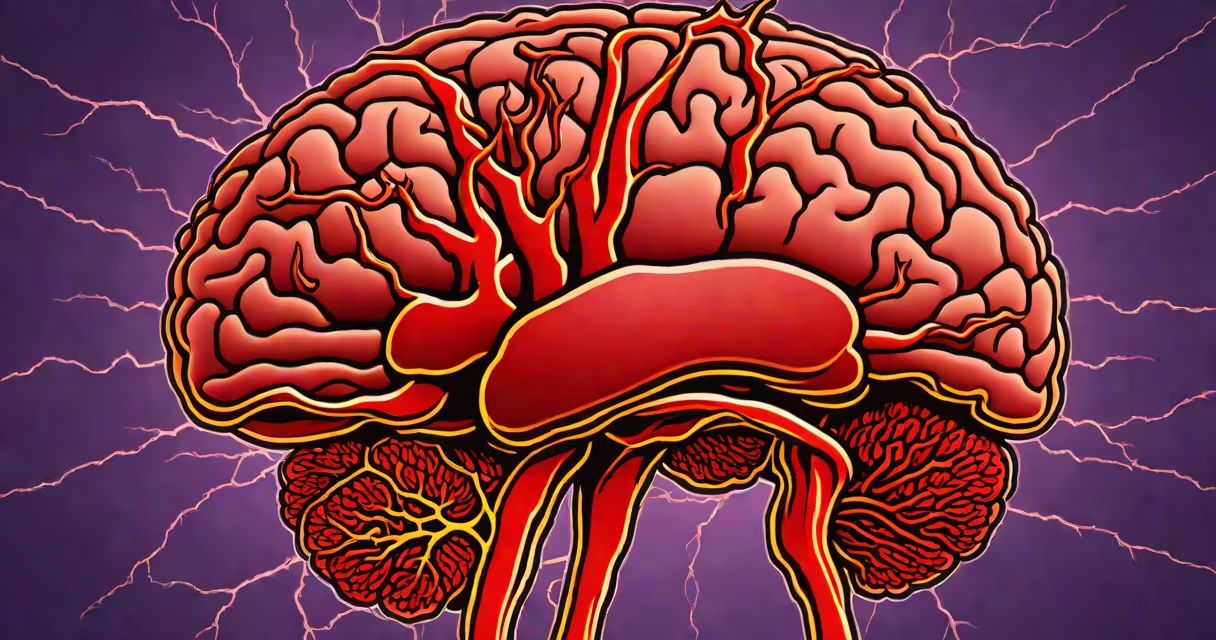
An embolic stroke happens when a blood clot forms in one part of the body-often the heart-and travels through the bloodstream until it lodges in a smaller artery in the brain, cutting off blood flow and causing tissue damage. This type of ischemic stroke accounts for about 20% of all ischemic events, according to the American Heart Association.
Unlike a thrombotic stroke, which forms directly in a brain artery, an embolic stroke originates outside the brain. The clot typically detaches from a site such as the left atrium in patients with atrial fibrillation, a condition affecting 2.7 million Americans. Once dislodged, the clot moves rapidly through the circulatory system. When it reaches a narrow cerebral vessel-usually in the middle cerebral artery-it blocks blood flow, leading to sudden neurological deficits.
Recovery depends heavily on time. For every minute untreated, 1.9 million neurons die, per research published in *The Lancet Neurology*. Prompt treatment with intravenous alteplase within 4.5 hours of symptom onset improves outcomes. Without timely intervention, permanent disability or death may follow.
Clots form due to abnormal blood flow or damaged vessel walls. In embolic stroke, the trigger is often cardiac arrhythmia, particularly atrial fibrillation. Atrial fibrillation increases the risk of clot formation in the left atrial appendage-a small pouch in the heart's upper chamber. These clots can remain attached for hours or days before breaking free.
Once released, the clot travels via the aortic arch into the carotid arteries, then into the brain's internal carotid arteries. The average clot size ranges from 2 to 6 millimeters. Because the brain's arteries branch into progressively smaller vessels, a clot of this size can easily obstruct a major cerebral artery.
When blood flow stops, oxygen delivery drops. Neurons begin dying within minutes. The area of dead tissue is called an infarct. Imaging scans-such as CT angiography or MRI diffusion-weighted imaging-can detect the location and extent of the damage within 30 minutes of arrival at a hospital.
Embolic strokes fall into distinct categories based on origin. The most common source is the heart, especially in people with structural heart disease or arrhythmias. Less frequent origins include large arteries in the neck or atherosclerotic plaques that break off during procedures.
| Source | Frequency | Typical Patient Profile | Prevention Strategy |
|---|---|---|---|
| Left atrial appendage (e.g., in AFib) | ~60% of embolic strokes | Aged 65+, history of hypertension or diabetes | Anticoagulants like apixaban or rivaroxaban |
| Carotid artery plaque | ~15% | History of transient ischemic attack (TIA) | Surgical endarterectomy or stenting |
| Heart valve disease | ~10% | Patient with prosthetic valves or infective endocarditis | Antibiotics and anticoagulation |
| Other (e.g., pulmonary embolism, rare) | <5% | Post-surgical or trauma-related | Prophylactic heparin during recovery |
One non-obvious fact: some emboli originate in the venous system and cross into the arterial circulation through a patent foramen ovale (PFO)-a small hole between the heart's upper chambers. This condition affects about 25% of adults. In rare cases, a clot from the leg veins can bypass the lungs and enter the brain, causing a paradoxical embolism.
Age, high blood pressure, diabetes, smoking, and high cholesterol increase the likelihood of developing conditions that lead to embolic stroke. But the strongest predictor is atrial fibrillation. People with untreated AFib have a fivefold higher risk of ischemic stroke compared to those without it, per the European Society of Cardiology guidelines.
Another overlooked factor is recent surgery. After orthopedic procedures-especially hip or knee replacement-patients face a temporary spike in clotting activity. The risk peaks around day 5 post-op. Anticoagulant therapy is standard during this window, but underuse remains a problem. A 2022 study in *JAMA Internal Medicine* found that only 68% of eligible patients received prophylaxis.
I once observed a 72-year-old man admitted with sudden aphasia and right-sided weakness. His EKG showed irregular atrial fibrillation. A CT scan revealed a large clot in the left middle cerebral artery. He received IV alteplase within 40 minutes of arrival. By day 3, his speech had improved. This case illustrates how quickly diagnosis and treatment can alter outcomes.
Embolic stroke is often confused with thrombotic stroke, but the difference lies in origin. Thrombotic strokes develop locally in a brain artery due to atherosclerosis. Embolic strokes come from distant sites. Another type, hemorrhagic stroke, results from bleeding into the brain and requires different treatment.
Key clinical clues help differentiate them. Embolic strokes tend to cause sudden onset of focal deficits-like facial droop or limb paralysis-without warning. They often affect the middle cerebral artery territory, leading to contralateral hemiparesis and aphasia. In contrast, thrombotic strokes usually progress over minutes to hours and may start with mild symptoms.
Imaging confirms the diagnosis. On non-contrast CT, early signs include loss of gray-white matter differentiation and hyperdense artery sign. MRI shows restricted diffusion in the affected region. Angiography reveals the occluded vessel and sometimes the source of the clot.
Recognizing symptoms early saves brain tissue. The FAST acronym-Face drooping, Arm weakness, Speech difficulty, Time to call emergency services-is recommended by the National Institute of Neurological Disorders and Stroke. If symptoms last longer than one minute, seek immediate care.
Prevention focuses on managing underlying conditions. Patients with atrial fibrillation should take anticoagulants unless contraindicated. Those with carotid stenosis greater than 70% may benefit from endarterectomy. Lifestyle changes-such as quitting smoking, reducing salt intake, and regular exercise-lower risk.
Understanding embolic stroke is critical because it's preventable in many cases. With proper screening, medication, and rapid response, outcomes improve dramatically. The goal is not just survival-but meaningful recovery.