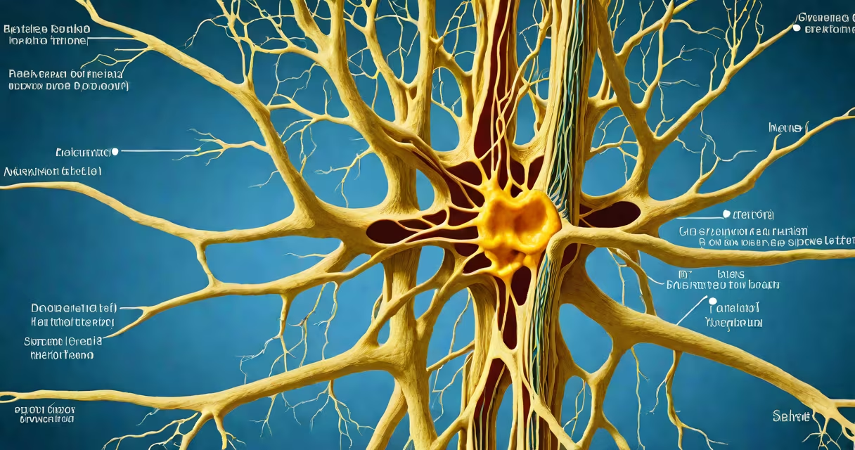
The components of the peripheral nervous system consist of nerves, sensory and motor neurons, and ganglia that relay signals between the central nervous system and the rest of the body. These structures enable voluntary movement, involuntary regulation of organs, and detection of environmental stimuli.
Nerves are bundles of axons extending from the spinal cord and brain to muscles, skin, and internal organs. They carry electrical impulses in both directions-sensory input toward the CNS and motor commands away from it. For example, the sciatic nerve, the longest in the body, transmits signals from the lower spine to the foot and leg, enabling walking and balance.
Sensory neurons detect changes in the environment-like temperature, pressure, or pain-and send that information to the central nervous system. Motor neurons transmit signals from the CNS to effectors such as muscles and glands. A single motor neuron can innervate hundreds of muscle fibers, allowing coordinated contraction during actions like lifting a cup.
The peripheral nervous system divides into two primary subsystems: the somatic nervous system and the autonomic nervous system. The somatic system controls voluntary movements and processes sensory input from the skin and joints. The autonomic system regulates involuntary functions such as heart rate, digestion, and respiratory rate.
The autonomic nervous system further splits into sympathetic and parasympathetic branches. Sympathetic activity prepares the body for stress-increasing heart rate, dilating pupils, and redirecting blood flow to muscles. Parasympathetic activity promotes calm states-slowing the heart, stimulating digestion, and conserving energy. This dual control allows precise adjustment of organ function based on context.
A key distinction often missed is that not all peripheral nerves contain both sensory and motor fibers. Some nerves are purely sensory (e.g., the optic nerve, which transmits visual data from the retina to the brain), while others are exclusively motor (e.g., the facial nerve's motor branch controlling facial expressions).
| Component | Type | Function | Example |
|---|---|---|---|
| Nerve | Bundle of axons | Transmits signals over distance | Radial nerve in arm |
| Sensory neuron | Unipolar or pseudounipolar | Captures external stimuli | Touch receptor in fingertip |
| Motor neuron | Bipolar or multipolar | Activates muscles/glands | Neuron in spinal cord controlling diaphragm |
| Ganglion | Cluster of neuron cell bodies | Processes signals outside CNS | Superior cervical ganglion regulating blood pressure |
Neurons in the peripheral nervous system have specialized structures. Sensory neurons typically have a single long dendrite and axon extending from the cell body, forming a U-shaped structure that allows rapid signal transmission. Motor neurons have multiple dendrites and a single axon projecting to their target tissue.
Ganglia are clusters of neuron cell bodies located outside the central nervous system. They serve as relay points for sensory information. For instance, the dorsal root ganglia contain the cell bodies of sensory neurons entering the spinal cord. Damage to these ganglia can result in loss of sensation or chronic neuropathic pain.
Peripheral nerves are protected by connective tissue sheaths: the endoneurium surrounds individual axons, the perineurium encases fascicles (bundles of axons), and the epineurium wraps the entire nerve. This layered protection prevents mechanical damage and maintains optimal conditions for signal conduction.
One widespread error is equating the peripheral nervous system with reflexes. While reflex arcs involve peripheral nerves, the processing occurs in the spinal cord-not in the PNS itself. Reflexes like pulling your hand from a hot stove happen quickly because the signal doesn't travel to the brain-synapses in the spinal cord trigger immediate motor responses.
Another confusion arises around the term "autonomic." People assume it means "automatic," but it specifically refers to involuntary control. The autonomic nervous system operates without conscious effort, yet it's not immune to influence. Mindfulness practices and biofeedback can modulate autonomic activity, such as reducing heart rate variability through controlled breathing.
During a clinical exam, doctors test peripheral nerve function using reflexes, sensation mapping, and muscle strength. For example, tapping the patellar tendon elicits a knee-jerk reflex mediated by the femoral nerve. Loss of this reflex may indicate damage to the L4 spinal nerve root or the peripheral nerve itself.
Consider a patient presenting with numbness in the left hand and weakness in finger movement. Physical examination reveals decreased sensation along the medial forearm and diminished grip strength. MRI shows no spinal cord lesion, but nerve conduction studies reveal slowed signal transmission in the ulnar nerve at the elbow.
This diagnosis points to cubital tunnel syndrome-a compression of the ulnar nerve as it passes behind the medial epicondyle of the humerus. The affected components of the peripheral nervous system include sensory axons carrying touch and pain signals from the ring and little fingers, and motor axons controlling intrinsic hand muscles. Treatment involves splinting, physical therapy, or surgery to relieve pressure.
Understanding the components of the peripheral nervous system clarifies how injuries, diseases, and treatments affect bodily function. For instance, diabetes can cause peripheral neuropathy due to prolonged high glucose damaging small sensory nerves. Early detection through routine screening improves outcomes.
Each component of the peripheral nervous system plays a distinct role in maintaining homeostasis and enabling interaction with the environment. Recognizing their structure and function supports accurate diagnosis, effective treatment, and informed health decisions.