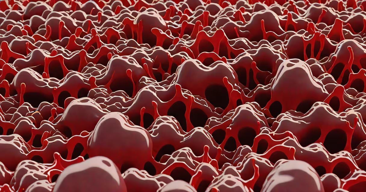
Blood clots form when platelets and proteins in plasma, especially fibrin, activate and bind together to stop bleeding after injury. This process is essential for wound healing but can become harmful if triggered inappropriately.
Blood clot formation begins when endothelial cells lining blood vessels are damaged. This exposes collagen and tissue factor, which initiate the coagulation cascade. Platelets adhere to the site within seconds, change shape, and release chemicals that attract more platelets. Within minutes, a fibrin mesh stabilizes the clot, sealing the breach.
The balance between clotting and bleeding is tightly regulated. Antithrombin, protein C, and tissue plasminogen activator (tPA) prevent excessive clotting. Disruption of this balance leads to either hemorrhage or thrombosis. The process is not random-it's a sequence of biochemical events triggered by specific physical and physiological cues.
Clotting occurs in three phases: vascular spasm, platelet plug formation, and coagulation. Vascular spasm reduces blood flow immediately after vessel injury. Platelets then aggregate at the site, forming a temporary plug. Finally, the coagulation cascade converts soluble fibrinogen into insoluble fibrin strands that reinforce the plug.
There are two pathways that trigger the coagulation cascade: intrinsic and extrinsic. The extrinsic pathway starts when tissue factor is released from damaged cells. The intrinsic pathway activates when blood contacts exposed collagen or other surfaces. Both converge on factor X, leading to thrombin generation and fibrin formation.
A single clot can form in under five minutes under normal conditions. For example, a paper cut typically results in a stable clot within three to four minutes. The process is localized-clotting remains confined to the injury site unless regulatory mechanisms fail.
Three elements drive clot formation: platelets, clotting factors, and fibrin. Platelets are cell fragments produced in bone marrow. They circulate in blood until activated by adhesion molecules like von Willebrand factor. Once activated, they release serotonin, ADP, and thromboxane A2, amplifying recruitment.
Coagulation factors are proteins synthesized in the liver. Factor VIII and IX deficiencies cause hemophilia. Vitamin K is required for the synthesis of factors II, VII, IX, and X. Without vitamin K, these factors remain inactive, increasing bleeding risk.
Fibrin forms the structural scaffold of a clot. Thrombin cleaves fibrinogen into fibrin monomers, which polymerize into long chains. These chains are cross-linked by factor XIIIa, creating a stable mesh. A single fibrin strand can be 10 micrometers long and withstand shear forces in flowing blood.
| Component | Source | Function | Deficiency Consequence |
|---|---|---|---|
| Platelets | Bone marrow | Adhere to injury site, release signaling molecules | Prolonged bleeding, petechiae |
| Fibrinogen | Liver | Converted to fibrin by thrombin | Impaired clot stability |
| Vitamin K | Diet, gut bacteria | Activates clotting factors II, VII, IX, X | Increased bleeding risk |
| Antithrombin | Liver | Inhibits thrombin and factor Xa | Hypercoagulable state |
Clots that form without injury or fail to dissolve are pathological. Deep vein thrombosis (DVT) occurs when clots develop in leg veins due to stasis, hypercoagulability, or endothelial damage-Virchow's triad. DVT affects about 1 in 1,000 people annually in the U.S., with higher rates in hospitalized patients.
Arterial clots often result from atherosclerotic plaques rupturing. Plaque erosion exposes collagen and tissue factor, triggering rapid platelet activation. An occlusive clot in a coronary artery causes myocardial infarction. About 805,000 Americans have a heart attack each year, with nearly half linked to sudden clot formation.
One non-obvious fact: clots can form in the absence of visible trauma. For example, prolonged immobility during long flights increases DVT risk by 30%. The mechanism is not mechanical injury but reduced venous return and altered blood flow patterns that promote platelet adhesion.
Many assume all clots are dangerous. In reality, most are protective. The body produces millions of clots daily to repair micro-injuries. Only when clots grow too large or form in the wrong location do they pose a threat.
Another misconception is that blood clots always cause pain. While clots in arteries may cause chest pain or limb ischemia, those in veins often go unnoticed until they dislodge and travel to the lungs-causing pulmonary embolism. Up to 10% of DVT cases lead to PE, and many are asymptomatic initially.
During menstruation, the uterine lining sheds. Blood loss exceeds 80 mL per cycle in about 10% of women. High estrogen levels delay endometrial breakdown, allowing blood to pool and clot. Clots larger than a quarter (2.5 cm) are abnormal and may signal fibroids or adenomyosis.
Menstrual clots are not always pathological. Some women naturally produce more fibrin due to genetic variation in factor XIII. However, frequent passage of large clots warrants evaluation for iron deficiency anemia or hormonal imbalance.
Anticoagulants like warfarin, rivaroxaban, and apixaban inhibit clotting factors. Warfarin blocks vitamin K-dependent factors, reducing thrombin generation. Rivaroxaban directly inhibits factor Xa. These drugs reduce stroke risk in atrial fibrillation by 60-70%.
Antiplatelet agents such as aspirin irreversibly inhibit cyclooxygenase-1, blocking thromboxane A2 production. Daily low-dose aspirin prevents recurrent heart attacks in high-risk individuals. The benefit is clear: it reduces major cardiovascular events by 20% over five years.
For patients with recurrent clots, testing for inherited disorders like factor V Leiden or prothrombin G20210A mutation is standard. These mutations increase clotting risk by 5-10 fold. Identification allows targeted prevention strategies.
Understanding what produces blood clots reveals a system designed for survival-but prone to error. The same mechanisms that seal wounds can obstruct blood flow. Recognizing early signs of abnormal clotting saves lives.