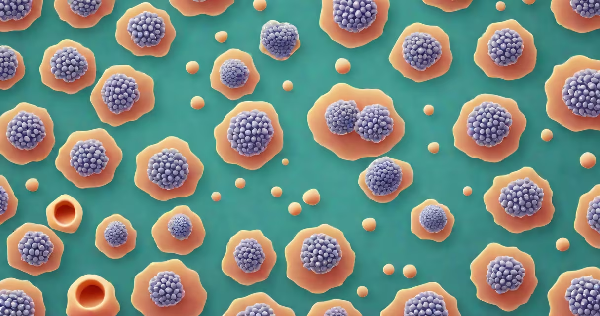
Dysplasia describes abnormal development or growth of cells in a tissue, often indicating a disruption in normal cellular maturation. It is not cancer but may signal early precancerous changes, especially when detected in epithelial linings such as the cervix, colon, or skin.
Dysplasia arises when cells lose their organized structure and begin dividing abnormally. This process typically occurs gradually, progressing through mild, moderate, and severe stages before potentially evolving into invasive cancer. The term originates from Greek roots: "dys" meaning bad or abnormal, and "plasis" meaning formation or shaping.
Pathologists identify dysplasia during biopsies using microscopic examination. Cells appear enlarged, irregular in shape, and show increased nuclear-to-cytoplasmic ratios. These features suggest disrupted differentiation and uncontrolled proliferation-hallmarks of early neoplastic change.
Dysplasia results from genetic or environmental stressors that interfere with normal cell cycle regulation. Common triggers include persistent viral infections (e.g., human papillomavirus in cervical dysplasia), chronic inflammation, or exposure to carcinogens like tobacco smoke.
Diagnosis begins with a physical exam or screening test-such as a Pap smear for cervical dysplasia or a colonoscopy for colonic lesions. If abnormal cells are seen, a biopsy confirms the presence and grade of dysplasia. The grading system ranges from low-grade (mild) to high-grade (severe), with high-grade dysplasia carrying a higher risk of progression to cancer.
For example, a 32-year-old woman with an abnormal Pap smear shows high-grade squamous intraepithelial lesion (HSIL)-a diagnosis equivalent to high-grade cervical dysplasia. A colposcopy-guided biopsy reveals cells with hyperchromatic nuclei, loss of polarity, and increased mitotic activity. This finding prompts immediate treatment to prevent progression to invasive carcinoma.
Dysplasia appears in various tissues, each with distinct clinical implications. The most common sites include:
| Tissue Type | Common Cause | Grading Scale | Clinical Implication |
|---|---|---|---|
| Cervix | HPV infection | Mild, Moderate, Severe | High-grade lesions may require loop electrosurgical excision (LEEP) |
| Colon | Chronic inflammation, polyps | Low-grade, High-grade | High-grade dysplasia in adenomas signals need for polypectomy |
| Lung | Tobacco use, asbestos | Grade I-IV | Often found in bronchial epithelium; linked to smoking-related cancer risk |
| Oral mucosa | Smokeless tobacco, alcohol | None, Mild, Moderate, Severe | Severe dysplasia requires biopsy and monitoring for oral cancer |
Each site uses slightly different classification systems. For instance, cervical dysplasia is graded using the Bethesda System, while colonic dysplasia follows the World Health Organization's classification. These systems standardize reporting and guide treatment decisions.
Dysplasia is frequently misunderstood as cancer. It is not. Dysplasia indicates a disturbance in cell maturation but lacks invasion beyond the basement membrane-a key criterion for malignancy. According to the American Cancer Society, less than 10% of high-grade dysplasia cases progress to invasive cancer within five years if monitored or treated appropriately.
Another misconception is that all dysplasia requires immediate intervention. That's false. Low-grade dysplasia in some organs-like the bladder or esophagus-may be monitored with regular follow-up rather than treated aggressively. The decision depends on location, severity, patient history, and risk factors.
A lesser-known fact: dysplasia can sometimes regress spontaneously. A 2019 study published in *The Lancet Oncology* found that 40% of women with low-grade cervical dysplasia experienced spontaneous resolution within two years without treatment. This underscores the importance of avoiding overtreatment in low-risk cases.
High-grade dysplasia generally warrants intervention due to its elevated risk of progression. Treatment methods depend on the affected organ. For cervical dysplasia, options include cryotherapy, LEEP, or cone biopsy. Colorectal dysplasia in a polyp is removed via endoscopic resection.
I once observed a 55-year-old man with high-grade dysplasia in a colon polyp during a routine screening. The polyp was removed endoscopically, and pathology confirmed complete excision with no residual dysplasia. He was advised to return for surveillance colonoscopy in three years-an outcome consistent with current guidelines from the American Gastroenterological Association.
Early detection remains critical. The National Institutes of Health reports that when high-grade dysplasia is identified and managed before invasion, five-year survival rates exceed 95%. This highlights the value of regular screening programs.
Dysplasia differs from metaplasia, which involves the replacement of one cell type with another-often as an adaptive response. Barrett's esophagus, for example, involves metaplasia of squamous epithelium into columnar type due to acid reflux. Over time, this metaplastic tissue may develop dysplasia, increasing cancer risk.
It also differs from hyperplasia, where cells increase in number but maintain normal architecture. Hyperplasia is reversible; dysplasia is not. A biopsy is required to distinguish between them.
Understanding what dysplasia means in medical terms enables informed discussions with healthcare providers. Recognizing it as a warning sign-not a diagnosis-helps patients make timely, evidence-based decisions about monitoring or treatment.