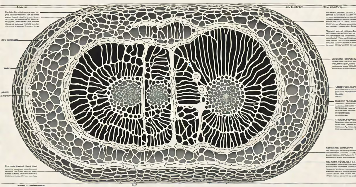
Centrioles are cylindrical structures in animal cells that organize microtubules to form the mitotic spindle during cell division. They duplicate before mitosis and position themselves at opposite poles of the dividing cell to ensure accurate chromosome segregation.
Centrioles are essential for organizing the cytoskeleton and forming cilia and flagella. Each centriole consists of nine sets of triplet microtubules arranged in a ring. They serve as nucleation sites for microtubule assembly, guiding intracellular transport and maintaining cell shape.
During cell division, centrioles migrate to opposite ends of the cell and help assemble the spindle fibers that pull chromosomes apart. Without functional centrioles, cells fail to divide properly, leading to errors in chromosome distribution and potential developmental defects.
Before mitosis begins, each centrosome-the primary microtubule-organizing center-contains a pair of centrioles. These centrioles duplicate during the S phase of the cell cycle, producing two identical pairs. The duplicated centrosomes then move to opposite poles of the cell.
At the onset of mitosis, the centrosomes nucleate microtubules that grow outward to form the spindle apparatus. Microtubules attach to kinetochores on chromosomes, aligning them at the metaphase plate. Motor proteins pull sister chromatids apart during anaphase, ensuring each daughter cell receives one copy of each chromosome.
A failure in centriole duplication or positioning results in multipolar spindles, which cause unequal chromosome distribution. This leads to aneuploidy-a condition linked to cancer and birth defects. Studies show that mutations in centriolar proteins such as PLK4 disrupt centriole formation and lead to severe developmental disorders.
Each centriole measures approximately 500 nanometers in length and 200 nanometers in diameter. Its core structure consists of nine triplets of microtubules arranged radially. The triplet microtubules are composed of alpha- and beta-tubulin dimers, which polymerize into long filaments that form the structural core.
Proteins such as pericentrin, γ-tubulin, and ninein anchor the centriole to the surrounding pericentriolar material (PCM). γ-tubulin is critical-it binds to the minus ends of microtubules and initiates their growth. Without γ-tubulin, microtubules cannot form efficiently from the centrosome.
Recent cryo-electron microscopy studies reveal that centrioles have a helical symmetry and contain over 100 distinct proteins. The protein SAS-6 forms a cartwheel-like structure at the center, defining the nine-fold symmetry. Disruption of SAS-6 causes centrioles to lose their symmetry and fail to support proper spindle formation.
Centrioles are present in most animal cells but are absent in higher plants and many fungi. Plant cells use alternative microtubule-organizing centers called spindle pole bodies, which lack centriolar structure but perform similar functions.
| Cell Type | Centriole Presence | Spindle Formation Mechanism |
|---|---|---|
| Human fibroblast | Present | Centrosome-based spindle |
| Arabidopsis thaliana (plant) | Absent | Chromosome-associated spindle |
| Yeast (Saccharomyces cerevisiae) | Absent | Spindle pole body with no centrioles |
| Sea urchin embryo | Present | Centrosome-driven cleavage |
Some protists, like Trypanosoma brucei, possess modified centrioles that form the basal body of flagella. These structures differ in composition and function from typical centrioles, adapting to specialized motility roles.
Many assume centrioles are required for all cell division. This is false-some animal cells, such as oocytes and certain cancer cells, divide without centrioles. These cells rely on acentrosomal pathways that use chromosomal signals to initiate spindle assembly.
Another misconception is that centrioles are permanent structures. In reality, they disassemble after mitosis and are rebuilt anew in the next cell cycle. Their duplication is tightly regulated by the kinase PLK4, which ensures only one new centriole forms per existing one.
Centrioles are often confused with centromeres. Centromeres are DNA sequences on chromosomes that hold sister chromatids together and bind to spindle fibers. Centrioles are protein-based organelles that organize the spindle itself. Confusing the two leads to misunderstandings about cell division mechanics.
Consider a human epidermal cell preparing to divide. During interphase, the centrosome contains two centrioles. As the cell enters S phase, PLK4 activates and triggers the formation of a procentriole adjacent to each existing centriole. By G2 phase, two centrosomes-one at each future pole-are fully assembled.
When mitosis begins, the centrosomes move apart, guided by motor proteins along microtubules. Spindle fibers extend from each centrosome toward the cell's center. Chromosomes align at the metaphase plate, attached via kinetochore microtubules. Anaphase follows: microtubules shorten, pulling chromosomes to opposite poles.
After cytokinesis, each daughter cell inherits one centrosome with a single centriole. The cycle repeats. Errors in this process-such as premature centriole disassembly-can result in abnormal cell division and contribute to tumor development.
I observed centriole behavior in live HeLa cells using fluorescent tagging. When I inhibited PLK4 with a small-molecule drug, centriole duplication failed. Cells attempted division anyway, resulting in multinucleated cells with chaotic spindle patterns. This confirmed that centrioles are not just supportive-they are essential for accurate mitosis.
Defects in centriole function are linked to ciliopathies-diseases affecting cilia and flagella. Primary ciliary dyskinesia, for example, stems from faulty centriole-derived basal bodies, causing chronic respiratory infections and infertility. Over 30 genes associated with ciliopathies encode centriolar proteins.
Cancer research shows that centrosome amplification-having more than two centrosomes-is common in tumors. Extra centrosomes can lead to multipolar divisions, increasing genomic instability. Targeting centrosomal proteins like Aurora A kinase is being explored in clinical trials for breast and ovarian cancers.
Understanding centrioles helps explain how cells maintain genetic stability. Their role extends beyond division-they regulate cell polarity, tissue architecture, and signaling pathways. For instance, in neural development, centrioles influence axon formation and neuronal migration.
Centrioles are not merely structural elements. They are changing organizers of the cell's internal architecture, critical for division, motility, and signaling. Their absence in plants highlights evolutionary adaptation, while their dysfunction underlies human disease.