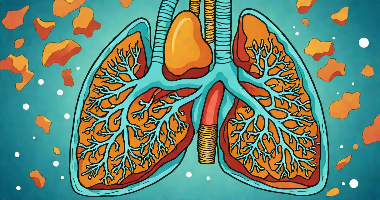
A collapsed lung, medically known as pneumothorax, happens when air accumulates in the pleural space-the area between the lung and the chest wall-causing the lung to lose its ability to expand and draw in oxygen. This condition can develop suddenly and requires prompt medical attention.
Pneumothorax disrupts normal breathing mechanics. The pleural space normally contains a small amount of fluid that allows the lung to glide smoothly against the chest wall during inhalation. When air enters this space due to a tear or rupture in the lung tissue or chest wall, pressure builds up and compresses the lung. Without intervention, the affected lung cannot fully inflate, leading to reduced oxygen intake and potential respiratory failure.
The most common cause is a spontaneous rupture of a small bleb-a blister-like growth-on the surface of the lung, especially in tall, thin young adults. Other triggers include trauma (such as rib fractures from a fall or car accident), underlying lung diseases like COPD or asthma, or complications from mechanical ventilation. A person may experience sudden sharp chest pain, shortness of breath, and a dry cough. In severe cases, the collapse can progress rapidly, requiring immediate emergency care.
The pleural cavity surrounds each lung and maintains negative pressure to keep the lungs inflated. When air enters this sealed space through a breach in the lung tissue or chest wall, the negative pressure is lost. As air accumulates, the lung collapses inward, reducing its volume and impairing gas exchange. The body responds with increased respiratory rate and effort to compensate for lower oxygen levels.
Diagnosis typically begins with a physical exam and confirmation via imaging. A standard chest X-ray shows the presence of air outside the lung and the visible retraction of lung tissue. In some cases, a CT scan provides higher detail, especially if the collapse is subtle or if there's suspicion of an underlying structural issue such as a cyst or tumor.
For example, a 24-year-old male with no history of lung disease presented with sudden left-sided chest pain and difficulty breathing after lifting heavy weights. A chest X-ray revealed a 30% left-sided pneumothorax. He was admitted for observation and treatment with supplemental oxygen and needle aspiration to remove the trapped air.
Pneumothorax is categorized by cause and severity. The main types are:
| Type | Cause | Typical Patient Profile | Treatment Approach |
|---|---|---|---|
| Primary Spontaneous | Rupture of subpleural blebs in healthy lungs | Young, tall, thin males (ages 15-35) | Oxygen therapy, observation; needle decompression if large |
| Secondary Spontaneous | Lung disease (e.g., emphysema, tuberculosis) | Older adults with chronic lung conditions | Often requires chest tube drainage |
| Traumatic | Blunt or penetrating injury (e.g., stab wound, fractured ribs) | Patients with recent trauma or surgery | Emergency chest tube placement; surgical repair if needed |
| tension | One-way valve effect trapping air under pressure | Life-threatening; can lead to cardiac arrest | Immediate needle decompression followed by tube thoracostomy |
Severity is measured by the percentage of lung collapse seen on imaging. A small pneumothorax (less than 20%) may resolve on its own with oxygen and monitoring. Larger collapses (over 30%) typically require drainage via a chest tube to remove the air and allow the lung to re-expand.
Many people confuse a collapsed lung with pneumonia or a pulmonary embolism. Pneumonia involves infection and inflammation of lung tissue, often with fever, productive cough, and abnormal lung sounds. A pulmonary embolism is a blood clot blocking a lung artery, presenting with sudden shortness of breath, chest pain that worsens with deep breaths, and sometimes hemoptysis (coughing up blood).
Another frequent error is assuming that a collapsed lung always results from trauma. While injuries are a major cause, spontaneous pneumothorax accounts for nearly half of all cases, particularly in otherwise healthy individuals. According to the American Thoracic Society, primary spontaneous pneumothorax occurs in about 7.4 per 100,000 people annually, mostly in men aged 20-40.
One non-obvious fact: recurrence rates are high. Up to 30% of patients who survive a first episode experience another collapse within five years. This risk increases if the initial event was large or if the patient has underlying lung disease. Preventive measures such as video-assisted thoracoscopic surgery (VATS) to remove blebs can reduce recurrence by over 90%.
Sudden onset of sharp, stabbing chest pain that worsens with breathing and is accompanied by shortness of breath is a red flag. If you or someone else experiences these symptoms, seek emergency care immediately. Delaying treatment increases the risk of progression to tension pneumothorax-a life-threatening condition where air continues to enter the pleural space but cannot escape, leading to mediastinal shift and compromised heart function.
I once observed a patient in the ER who had a 50% right-sided pneumothorax after a minor fall. He initially dismissed his symptoms as muscle strain. By the time he arrived at the hospital, his oxygen saturation had dropped to 88% on room air. After chest tube insertion, his lung re-expanded within hours, and he recovered fully. This case underscores that even mild trauma can trigger a serious event in susceptible individuals.
Not all collapses require hospitalization. Small, stable pneumothoraces in low-risk patients may be managed outpatient with close follow-up. However, any suspected case should be evaluated by a healthcare provider. Treatment depends on size, symptoms, and patient health status.
Understanding what it means if your lung collapses is essential for recognizing early warning signs and seeking appropriate care. Prompt diagnosis and treatment prevent complications and improve recovery outcomes. Recognizing the difference between a collapsed lung and similar conditions ensures faster access to effective interventions.