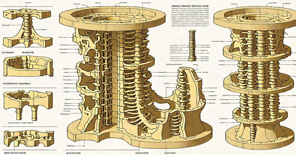
The vertebral column, also known as the spine or spinal column, is a flexible, segmented structure of 33 individual bones called vertebrae that form the core of the human axial skeleton. It encases and protects the spinal cord, supports the head and trunk, and enables upright posture and movement.
Each vertebra consists of a body, a neural arch, and processes that articulate with adjacent vertebrae. These bones are stacked vertically, forming a hollow tube through which the spinal cord passes. Intervertebral discs-fibrocartilaginous pads between the bodies-absorb shock and allow limited motion between segments.
The vertebral column is divided into five regions: cervical (7 vertebrae), thoracic (12), lumbar (5), sacral (5 fused into one sacrum), and coccygeal (3-4 fused into the coccyx). This segmentation allows for specialized functions: cervical vertebrae enable neck rotation, thoracic vertebrae stabilize the rib cage, and lumbar vertebrae bear most of the body's weight.
Movement in the vertebral column occurs through controlled articulation at synovial joints between adjacent vertebrae. The intervertebral discs act as cushions, distributing compressive forces during walking, lifting, or bending. The ligaments-such as the anterior and posterior longitudinal ligaments-stabilize the column and prevent excessive motion.
Nerve roots exit the spinal cord through intervertebral foramina, small openings between adjacent vertebrae. Compression of these roots due to disc herniation or spinal stenosis can cause pain, numbness, or weakness in limbs. For example, a herniated disc at L4-L5 may compress the L5 nerve root, leading to radiating pain down the leg, a condition known as sciatica.
Posture relies heavily on the vertebral column's natural curves. The spine has four primary curvatures: cervical (lordotic), thoracic (kyphotic), lumbar (lordotic), and sacral (kyphotic). These curves develop during infancy and early childhood as the child learns to sit, stand, and walk. Without proper development, conditions like scoliosis (lateral curvature) or kyphosis (excessive forward curve) can emerge.
Vertebrae differ across regions. Cervical vertebrae have transverse foramina for vertebral artery passage. Thoracic vertebrae feature costal facets for rib attachment. Lumbar vertebrae are larger and more solid to withstand mechanical loads. The sacrum forms a rigid base of the pelvis, fusing during adolescence.
| Region | Number of Vertebrae | Key Features | Primary Function |
|---|---|---|---|
| Cervical | 7 | Transverse foramina, bifid spinous processes | Head movement, support |
| Thoracic | 12 | Rib facets, long spinous processes | Thoracic cage stability |
| Lumbar | 5 | Large bodies, short blunt processes | Weight-bearing, trunk flexion |
| Sacral | 5 fused | Ala, sacral hiatus | Pelvic support, nerve exit points |
| Coccygeal | 3-4 fused | Small, rudimentary | Support during sitting |
A common misconception is that the vertebral column is a single rigid rod. In reality, it is highly adaptable. The cervical region allows up to 80 degrees of rotation, while the lumbar spine permits about 40 degrees of flexion. These ranges are essential for daily activities such as turning to look behind or bending to tie shoes.
Many assume the vertebral column is solely responsible for structural support. While it provides the main structure, much of the load is shared with the pelvic girdle and muscles. The erector spinae and abdominal muscles work synergistically with the spine to maintain balance and reduce injury risk.
Another error is equating back pain with spinal degeneration. Not all back pain stems from vertebral abnormalities. A 2021 study published in JAMA Internal Medicine found that 85% of low back pain cases have no identifiable structural cause on imaging. Pain often arises from muscle strain, poor biomechanics, or nerve irritation-not bone or disc pathology.
The vertebral column does not grow in length after puberty. Growth occurs primarily in the intervertebral discs during childhood. By age 16, the spine reaches approximately 90% of adult height. After that, changes in height result from disc compression, not bone growth.
In my experience as a medical educator, students often struggle to visualize the vertebral column's three-dimensional orientation. During dissection labs, I've seen learners misidentify the cervical vertebrae because they expect all vertebrae to resemble the lumbar ones. The atlas (C1) lacks a body and has a ring-like structure, allowing the head to nod "yes." The axis (C2) features the dens, a tooth-like projection that enables rotational movement.
Consider a patient presenting with neck stiffness and headache. Imaging reveals a fracture of the C1 ring. Because the vertebral artery passes through the transverse foramen of C1, trauma here risks arterial injury, potentially causing stroke. Immediate immobilization and neurovascular monitoring are critical-this is not a minor injury.
When evaluating spinal alignment, clinicians use the Cobb angle to measure scoliosis severity. A curve exceeding 10 degrees is diagnosed as scoliosis. Curves above 45 degrees may require surgical intervention. This metric underscores how precise measurements define clinical decisions.
The vertebral column is not merely a passive scaffold. It is a changing structure enabling locomotion, protecting delicate neural tissue, and adapting to mechanical stress. Its design reflects millions of years of evolutionary optimization for bipedalism, balance, and resilience under variable loads.
Understanding the vertebral column is essential for diagnosing musculoskeletal disorders, planning orthopedic interventions, and promoting spinal health. Whether assessing posture, treating injuries, or studying anatomy, the vertebral column remains central to human function and well-being.
For context on how the spine connects to the skull and supports the entire axial skeleton, explore the relationship between the skull and spinal column.