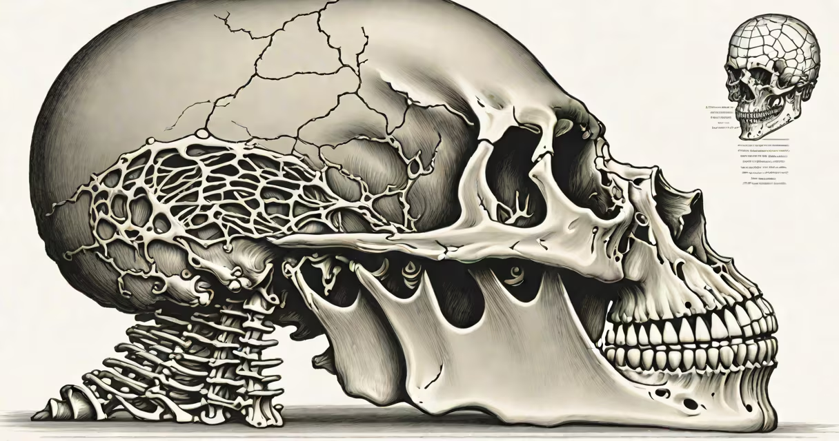
The skull and spinal column are the primary structural components of the human axial skeleton, forming a continuous bony enclosure that protects the brain and spinal cord-the central nervous system. Together, they provide mechanical support, house critical neural tissue, and serve as attachment points for muscles and ligaments.
These structures begin at the base of the skull, where the occipital bone articulates with the first cervical vertebra (C1), known as the atlas. This joint allows for nodding movements, such as saying "yes." From there, the spinal column extends downward through seven cervical, twelve thoracic, and five lumbar vertebrae, followed by the sacrum and coccyx. The skull encloses the cranial cavity, which contains the brain, while the vertebral canal formed by the spinal column houses the spinal cord.
Each segment of the spinal column is separated by intervertebral discs-fibrocartilaginous pads that absorb shock and allow flexibility. The skull's bones are fused at sutures, except at the temporomandibular joint, which enables jaw movement. These joints and articulations allow for precise motion while maintaining protection for delicate neural tissues.
The skull and spinal column function as a rigid yet adaptable scaffold. The skull's thick, dense bones-such as the frontal, parietal, and temporal bones-resist impact and prevent deformation of the brain during trauma. The dura mater, a tough outer membrane of the meninges, lines the inner surface of the skull and further cushions the brain.
Inside the spinal column, the spinal cord runs from the medulla oblongata (part of the brainstem) down to the level of the first lumbar vertebra (L1). Nerve roots exit between each vertebra, forming peripheral nerves that control sensation and motor function throughout the body. For example, damage to the C5-C6 region can result in weakness in the biceps and wrist extensors.
A single misalignment in the cervical spine-such as a herniated disc at C6-can compress a nerve root, causing pain radiating into the thumb side of the hand. This illustrates how structural integrity directly affects neurological function. The spinal column's role in signal transmission is not passive; it actively modulates sensory input and motor output via reflex arcs and ascending/descending tracts.
The skull consists of eight cranial bones and fourteen facial bones. The cranial bones include the frontal, two parietal, two temporal, occipital, sphenoid, and ethmoid bones. They fuse during development, with the anterior fontanelle closing around 18 months of age. The facial bones form the structure of the face and support the eyes, nose, and mouth.
The spinal column is divided into five regions based on location and function:
| Region | Number of Vertebrae | Key Functions |
|---|---|---|
| Cervical | 7 | Supports head, allows rotation and flexion |
| Thoracic | 12 | Attaches to ribs; limits mobility for chest protection |
| Lumbar | 5 | Bears most body weight; enables bending and lifting |
| Sacral | 5 fused | Forms pelvic floor; supports abdominal organs |
| Coccygeal | 3-4 fused | Seat support; minimal movement |
Each vertebra has a body, pedicles, laminae, and processes. The vertebral foramen forms the vertebral canal. The spinal cord occupies this space from the foramen magnum to L1. Below that, the cauda equina-a bundle of spinal nerves-extends downward like a horse's tail.
Many assume the spine is a single solid rod. In reality, it has four natural curves: cervical (lordotic), thoracic (kyphotic), lumbar (lordotic), and sacral (kyphotic). These curves improve balance and reduce stress during walking and standing.
Another misconception is that the skull is entirely immovable. While the bones are fused in adults, subtle movements occur during respiration and blood flow. The cranial bones are connected by fibrous sutures that allow minute expansion and contraction-measured clinically in intracranial pressure monitoring.
The skull and spinal column do not operate independently. The brainstem, located at the junction of the brain and spinal cord, regulates important functions like breathing and heart rate. A fracture at the base of the skull can disrupt this area, leading to life-threatening complications. For example, a basilar skull fracture may present with raccoon eyes (periorbital ecchymosis) or Battle's sign (bruising behind the ear).
In emergency medicine, the presence of a skull fracture increases the risk of intracranial hemorrhage. A CT scan showing a linear fracture near the middle meningeal artery requires immediate evaluation due to the potential for epidural hematoma. This condition can progress rapidly, requiring surgical evacuation within hours.
Spinal injuries are similarly high-stakes. A compression fracture of the T12 vertebra can result from a fall from height. In one documented case, a 42-year-old man fell 12 feet from a ladder and sustained a T12 burst fracture. Imaging revealed spinal cord compression, necessitating surgical stabilization with pedicle screws and rods. Post-op MRI confirmed decompression of the cord.
During a routine physical exam, a physician assesses spinal alignment using the Adam's forward bend test to detect scoliosis. The patient bends forward at the waist, and the examiner checks for asymmetry in the rib cage or shoulder height. A difference of more than 5 degrees indicates a need for further imaging.
I once observed a patient with chronic neck pain who had developed a C5-C6 disc herniation. After six weeks of conservative treatment failed, an MRI showed significant spinal cord compression. Surgical discectomy and fusion restored function. The patient regained full strength in the biceps and returned to work within three months.
The skull and spinal column are not merely protective shells-they are integrated systems. Nerves exiting the spinal cord travel through intervertebral foramina, while cranial nerves pass through bony foramina in the skull. Damage to either structure can cause paralysis, sensory loss, or autonomic dysfunction.
For instance, a tumor pressing on the medulla can impair respiratory rhythm. Similarly, a fracture of the clivus-a bony region at the base of the skull-can affect cranial nerves III, IV, V, and VI, leading to double vision, facial numbness, and difficulty swallowing.
These structures represent both resilience and vulnerability. Their design balances protection with mobility, but their complexity makes them susceptible to injury and disease. Recognizing their role in health and illness improves outcomes in trauma, degenerative disorders, and congenital anomalies.