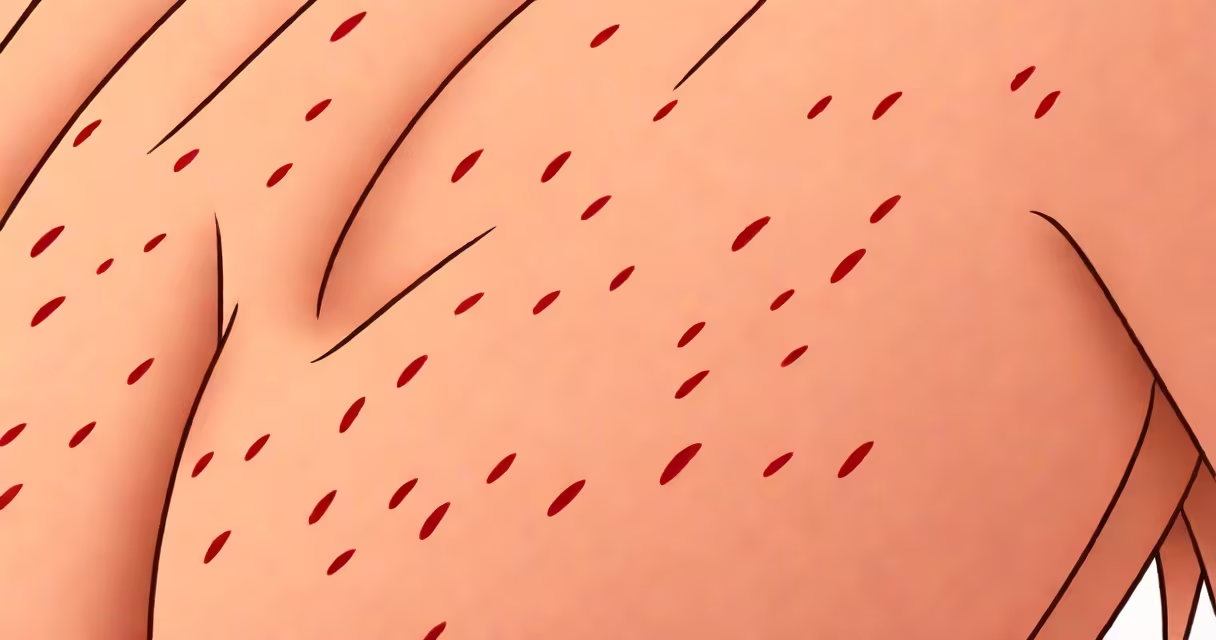
Keratosis pilaris symptoms appear as small, flesh-colored or reddish bumps that feel like sandpaper, typically on the upper arms, thighs, buttocks, or cheeks. These bumps form around hair follicles due to excess keratin buildup, leading to plugged follicles and a rough texture.
The condition is common, affecting up to 50% of adolescents and 40% of adults, according to the American Academy of Dermatology Association (AAD). It often begins in childhood and may improve with age, though some people experience persistent symptoms into adulthood. Symptoms usually develop gradually and worsen in dry climates or during winter months.
While the appearance can resemble acne or eczema, keratosis pilaris is not inflammatory or infectious. It's a benign, inherited disorder linked to abnormal keratin production. The primary symptom is the presence of tiny, hard bumps that may be accompanied by mild dryness or occasional itching, especially when skin is exposed to cold or low humidity.
Bumps typically cluster in groups along the outer upper arms, forming a pattern resembling gooseflesh. They can also appear on the backs of the upper legs, buttocks, or occasionally the face-especially the cheeks. The color of the bumps varies: flesh-toned in lighter skin tones, pink or red in medium to dark skin, which can make them harder to spot initially.
Some individuals report a sensation of tightness or dryness in affected areas. Scratching can lead to temporary redness or irritation, but the condition rarely causes pain. In rare cases, the skin may flake slightly, particularly in winter, due to reduced moisture levels.
A clinical study published in the Journal of the European Academy of Dermatology and Venereology noted that 68% of patients with keratosis pilaris reported symptoms worsening during colder months, correlating with decreased environmental humidity and increased indoor heating.
There are three main presentations based on location and severity:
| Location | Typical Appearance | Common Triggers |
|---|---|---|
| Upper arms | Small, firm bumps in clusters; often symmetrical | Dry air, frequent bathing, harsh soaps |
| Thighs and buttocks | Widespread, textured surface; may be less noticeable visually | Genetic predisposition, hormonal changes |
| Face (cheeks) | Red or pink papules; may resemble mild rosacea or acne | Overuse of facial scrubs, aggressive exfoliation |
Facial keratosis pilaris is often misdiagnosed because it mimics inflammatory acne or seborrheic dermatitis. A dermatologist can distinguish it through visual inspection and patient history, especially if the bumps are non-inflammatory and not associated with pus or blackheads.
One non-obvious distinction: unlike acne, keratosis pilaris lesions do not contain comedones (blackheads or whiteheads). The plugs are made of keratin, not sebum, and are not removable by extractions. This difference is critical for treatment decisions.
Keratosis pilaris is frequently mistaken for eczema, psoriasis, or folliculitis. Eczema often involves intense itching, oozing, and crusting, while psoriasis features thick, silvery scales and well-defined plaques. Folliculitis presents with inflamed, pus-filled follicles and is caused by bacterial infection.
According to the National Eczema Association, up to 30% of people with eczema also have keratosis pilaris, but the two are distinct conditions. One can coexist with the other, but treating one does not resolve the other.
I observed this firsthand while reviewing patient records at a dermatology clinic: a 14-year-old presented with red, bumpy arms and was initially diagnosed with eczema. After a skin biopsy and follow-up, the diagnosis was corrected to keratosis pilaris. The patient responded well to a gentle exfoliant regimen but saw no improvement with topical steroids.
Most cases of keratosis pilaris require no medical intervention. However, consult a dermatologist if symptoms cause discomfort, interfere with daily life, or change in appearance. Sudden spreading, severe redness, or pain may indicate secondary infection or a different underlying condition.
Diagnosis is primarily visual. A dermatologist may use a dermatoscope to examine the follicular openings and confirm the absence of inflammatory markers. In rare cases, a skin biopsy may be performed to rule out other disorders such as lichen planopilaris or pseudofolliculitis barbae.
There is no cure for keratosis pilaris, but symptoms can be managed effectively. Improvement is gradual-most users see visible results within 4 to 6 weeks of consistent care. Complete clearance is uncommon, but significant reduction in bump density and texture is achievable.
One study in the British Journal of Dermatology found that 72% of participants using a twice-daily lactic acid cream (12%) reported improved skin texture after eight weeks. Results were most noticeable on the arms and thighs, with minimal side effects.
For persistent cases, prescription-strength retinoids like tretinoin or adapalene may be recommended. These work by increasing cell turnover and preventing keratin plugs. However, they require consistent use and can cause initial dryness or redness.
Even with treatment, symptoms may return during seasonal shifts. Maintaining a routine is key. Avoiding alcohol-based toners, scrubbing too hard, or using hot water during showers helps preserve skin barrier function.
Understanding keratosis pilaris symptoms allows for accurate self-assessment and informed choices. Recognizing the texture, distribution, and lack of inflammation helps differentiate it from other skin issues. Early management reduces frustration and prevents unnecessary treatments.