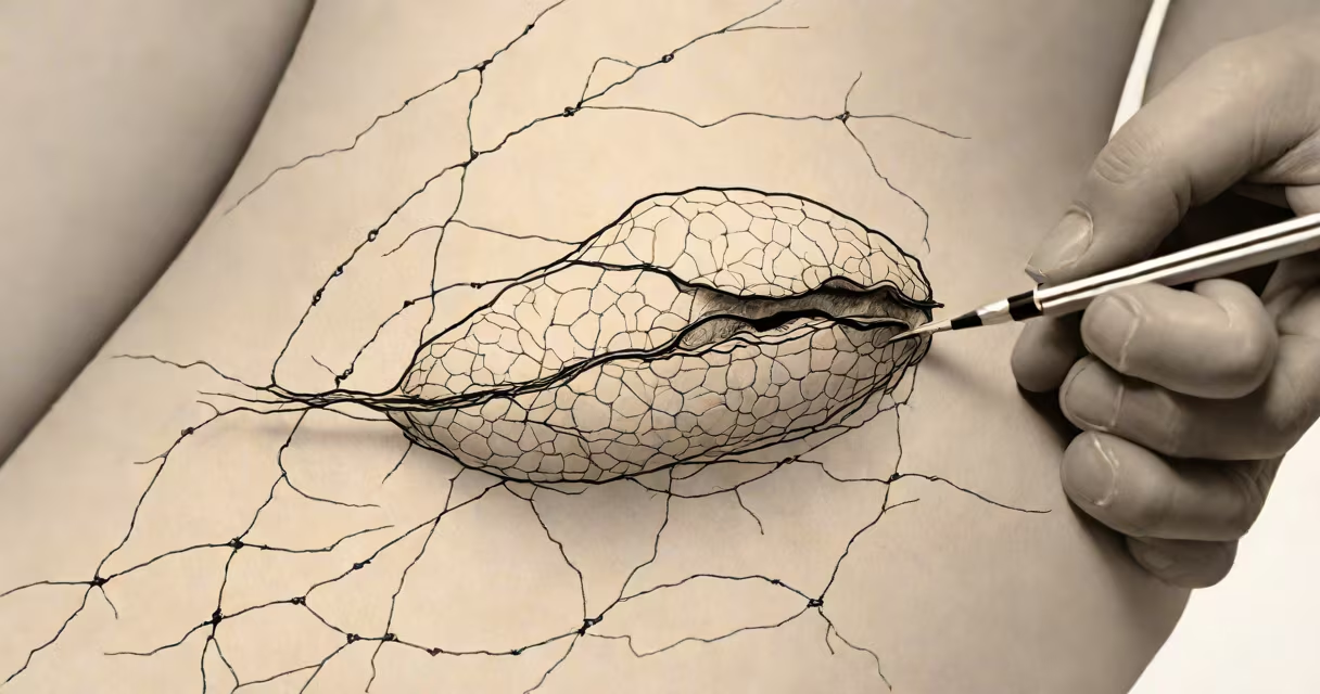
A suture granuloma is a chronic inflammatory lesion formed around a non-absorbable suture material left in the body after surgery. It develops when immune cells surround the suture as a foreign body, leading to localized swelling, pain, or drainage. This condition typically appears weeks to months after surgery, often mimicking infection or recurrence of disease.
Suture granulomas arise when the body fails to fully absorb or tolerate synthetic or non-resorbable suture materials such as polypropylene, polyester, or nylon. These materials persist in soft tissues and trigger a persistent immune response. The granuloma forms as macrophages and multinucleated giant cells gather around the suture filament, attempting to wall off the foreign substance. Histologically, this results in a well-circumscribed nodule composed of epithelioid cells and fibrous tissue.
According to the American Journal of Clinical Pathology, suture granulomas account for approximately 1-3% of all cutaneous granulomatous lesions seen in dermatopathology practice. They are most common in areas where sutures are frequently used-abdominal, breast, and facial surgeries-but can occur anywhere sutures were placed.
The process begins when a non-absorbable suture is implanted during surgery. Unlike absorbable sutures (e.g., Vicryl or Monocryl), which degrade over time, non-absorbable sutures remain intact indefinitely. The body recognizes them as foreign and mounts a delayed-type hypersensitivity reaction. Immune cells infiltrate the area, releasing cytokines that attract more macrophages.
Over time, macrophages fuse into multinucleated giant cells, forming the core of the granuloma. This leads to fibrosis and tissue remodeling. The lesion may appear as a painless or tender nodule, sometimes draining serosanguinous fluid. In rare cases, it can erode through the skin surface, creating an open wound that resembles a chronic ulcer.
One key factor influencing development is suture size and material. A 2018 study in the Journal of Surgical Research found that larger-diameter sutures (e.g., 3-0 or 4-0 polypropylene) were associated with a higher incidence of granuloma formation compared to finer threads. The surface area of the suture correlates with the magnitude of the foreign body reaction.
Suture granulomas most frequently develop at sites of prior surgical incisions. The most common locations include:
Clinically, patients report a slowly enlarging lump under the skin, often near the original scar. The lesion may be firm, mobile, and occasionally painful. Skin changes include redness, warmth, or breakdown with drainage. Symptoms typically emerge between 6 weeks and 18 months post-op, though cases have been documented as late as 5 years after surgery.
For example, a 52-year-old woman developed a 1.5 cm nodule at her right lower quadrant incision site 11 months after a laparoscopic cholecystectomy. She had no fever or systemic symptoms. Ultrasound showed a hypoechoic mass with internal vascularity. Biopsy confirmed a granulomatous reaction surrounding a polypropylene suture fragment.
Suture granulomas are often mistaken for recurrent cancer, abscesses, or infections. Their appearance overlaps with other granulomatous disorders such as sarcoidosis or tuberculosis, especially on imaging. However, key differences exist.
Unlike malignancy, suture granulomas do not show cellular atypia or rapid growth. They lack necrotizing features typical of tuberculous granulomas. The presence of a known surgical history and a suture fragment on histology confirms the diagnosis. Imaging may show a well-defined mass with peripheral enhancement, but no lymphadenopathy or distant spread.
A critical distinction lies in the timing. Infections usually present within days to weeks post-op with fever, purulent discharge, and acute inflammation. Suture granulomas appear later, with a subacute course. This delay is due to the delayed immune response to the foreign material.
One non-obvious fact: suture granulomas can form even when the suture was buried deep in tissue. The immune system can detect the foreign body regardless of depth. A 2020 case report in the Annals of Plastic Surgery described a granuloma beneath the dermis of a reconstructed breast flap, visible only via MRI and confirmed by excisional biopsy.
Diagnosis relies on clinical suspicion, imaging, and definitive histopathological analysis. Fine-needle aspiration (FNA) may suggest granulomatous inflammation but cannot confirm suture origin. Definitive diagnosis requires tissue sampling-either punch biopsy or excision.
Imaging modalities such as ultrasound or MRI can identify a mass with a linear or thread-like structure suggestive of a suture. However, these tools cannot distinguish between a suture and a retained instrument fragment. Only pathology can confirm the presence of synthetic fibers.
Management depends on severity. Asymptomatic granulomas may be observed. Painful or draining lesions require surgical excision. Complete removal of the suture material is essential to prevent recurrence. Incomplete excision leaves residual material, leading to persistent inflammation.
I once observed a patient with a recurrent granuloma at a cesarean section scar. After two failed biopsies, we performed wide local excision with removal of the entire suture tract. The lesion did not return over 18 months of follow-up. This underscores the importance of complete excision.
Treatment options include:
| Approach | When Used | Success Rate |
|---|---|---|
| Observation | Asymptomatic, small nodules | ~60% |
| Punch biopsy | Diagnostic confirmation | ~85% |
| Excisional surgery | Painful, draining, or growing lesions | ~95% |
Non-surgical treatments like corticosteroid injections have limited success. A 2019 study in Dermatologic Surgery reported a 40% resolution rate with intralesional triamcinolone, but relapse occurred in 25% of cases within six months.
Preventing suture granulomas involves selecting appropriate suture materials based on tissue type and location. For superficial wounds, absorbable sutures reduce long-term risk. In deeper layers, using fine-gauge non-absorbable sutures minimizes foreign body burden.
Surgeons should avoid leaving excess suture ends in tissue. All suture tails should be trimmed flush with the skin or fascia. Some institutions now mandate suture documentation in operative notes, including material type and length used.
Patients should be informed about potential late complications. A 2021 review in the British Journal of Surgery emphasized that patient education reduces unnecessary anxiety and improves early reporting of unusual symptoms.
Understanding what is a suture granuloma enables accurate diagnosis and timely intervention. It is not a sign of infection or cancer, but a predictable biological response to persistent foreign material. Early recognition prevents misdiagnosis and avoids prolonged treatment delays.