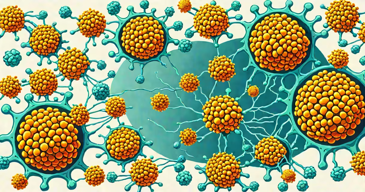
T cells are a type of white blood cell central to the adaptive immune system, responsible for identifying and eliminating infected, damaged, or cancerous cells through targeted attacks. They mature in the thymus and coordinate immune responses by recognizing specific antigens presented by other cells.
These cells form the core of cell-mediated immunity, distinguishing them from antibody-producing B cells. They do not neutralize pathogens directly but activate other immune cells or destroy compromised cells themselves. Their function is essential for long-term immunity and immune memory.
T cells detect foreign or abnormal proteins displayed on the surface of host cells via major histocompatibility complex (MHC) molecules. MHC class I presents antigens from intracellular pathogens like viruses, while MHC class II presents antigens from extracellular threats such as bacteria.
When a T cell receptor binds to an antigen-MHC complex, the T cell becomes activated. This triggers proliferation and differentiation into effector cells. For example, cytotoxic T cells release perforin and granzymes to induce apoptosis in infected cells, effectively killing them without harming surrounding tissue.
A single activated T cell can generate thousands of daughter cells within days. This clonal expansion ensures a rapid response during reinfection, forming the basis of immunological memory. Memory T cells persist for years, enabling faster reactions upon re-exposure to the same pathogen.
There are several functional subtypes of T cells, each with distinct responsibilities:
| Type | Function | Activation Signal | Target |
|---|---|---|---|
| Cytotoxic T cells (CD8+) | Directly kill virus-infected or cancerous cells | Antigen + MHC class I | Infected cells, tumor cells |
| Helper T cells (CD4+) | Coordinate immune responses by releasing cytokines | Antigen + MHC class II | B cells, macrophages, other T cells |
| Regulatory T cells (Tregs) | Suppress immune activity to prevent autoimmunity | Antigen + MHC class II + co-stimulatory signals | Self-reactive T cells, inflammatory sites |
CD4+ helper T cells are further divided into subsets based on cytokine profiles and transcription factors. Th1 cells promote cellular immunity against intracellular pathogens, while Th2 cells support humoral immunity by aiding B cell antibody production. Th17 cells drive inflammation in mucosal defenses, and T follicular helper (Tfh) cells assist B cells in germinal centers to produce high-affinity antibodies.
Regulatory T cells maintain tolerance to self-antigens. Without them, the immune system attacks healthy tissues, leading to autoimmune diseases like type 1 diabetes or multiple sclerosis. Studies show that mutations in the FOXP3 gene disrupt Treg development, resulting in severe autoimmunity in humans (Nature Immunology, 2021).
T cells operate primarily in secondary lymphoid organs-lymph nodes, spleen, and mucosal tissues-where they encounter antigen-presenting cells like dendritic cells. These cells process and display fragments of pathogens, triggering T cell activation.
Once activated, T cells migrate to infection sites. Cytotoxic T cells patrol tissues, scanning for cells displaying viral peptides. Upon detection, they bind via T cell receptors and deliver lethal hits. A single cytotoxic T cell can eliminate up to 100 infected cells per day.
Helper T cells amplify immune responses by secreting signaling molecules. Interleukin-2 (IL-2) promotes T cell growth; interferon-gamma (IFN-γ) activates macrophages. These signals ensure coordinated action across immune cell types.
One non-obvious fact: T cells can recognize antigens derived from intracellular proteins even when those proteins are not expressed on the cell surface. This happens through proteasomal degradation and transport of peptide fragments into the endoplasmic reticulum, where they load onto MHC class I molecules.
T cells are often confused with B cells, which produce antibodies. While both are lymphocytes, they differ fundamentally in mechanism. B cells respond to free-floating antigens and generate humoral immunity, whereas T cells require antigen presentation on cell surfaces and mediate cell-mediated immunity.
Another misconception is that T cells attack all foreign invaders. In reality, they only respond to specific antigen-MHC complexes. This specificity prevents collateral damage but requires precise antigen processing and presentation.
Some assume T cells act independently. In practice, they rely on cross-talk with dendritic cells, macrophages, and B cells. For example, a helper T cell must receive co-stimulation (e.g., CD28-B7 interaction) to become fully activated-otherwise, it may become anergic (unresponsive).
I observed this during a lab experiment using flow cytometry: T cells exposed to antigen without co-stimulation showed no proliferation, confirming that signal 2 is essential for full activation.
Consider a person infected with influenza. Dendritic cells in the lungs capture viral proteins, migrate to draining lymph nodes, and present viral peptides on MHC class I and II molecules.
CD8+ T cells recognize viral peptides on MHC I and differentiate into cytotoxic effectors. Within five days, they infiltrate lung tissue, locate infected epithelial cells, and induce apoptosis. This reduces viral replication and limits disease spread.
Simultaneously, CD4+ helper T cells recognize antigens on MHC II and secrete IL-2 and IFN-γ. These signals boost cytotoxic T cell activity and improve macrophage function. After recovery, memory T cells remain in circulation, ready to respond rapidly if the same strain reappears.
This coordinated effort highlights why T cells are critical for controlling infections that evade antibody-based defenses, such as HIV, which mutates rapidly and hides inside host cells.
T cells are indispensable in defending the body against intracellular threats, maintaining immune balance, and enabling lasting protection. Their role extends beyond infection control-they also monitor for cancerous changes and regulate immune tolerance.