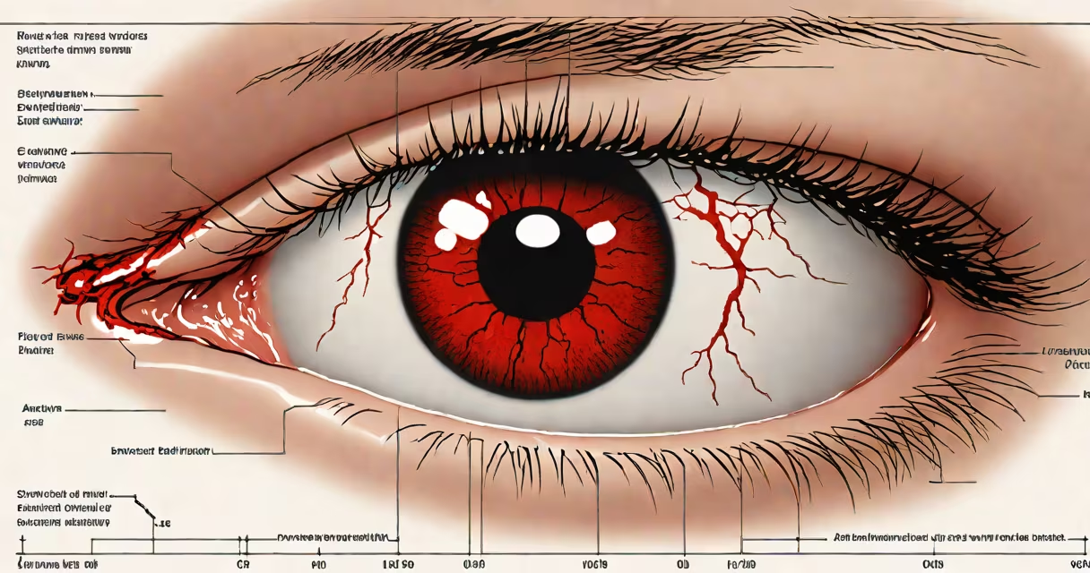
A red blood spot in the white part of the eye, medically known as a subconjunctival hemorrhage, occurs when a tiny blood vessel under the clear surface of the eye bursts. It appears as a vivid red or pink patch on the sclera and often causes concern despite being painless and typically resolving on its own.
This condition results from minor trauma, sudden increases in blood pressure, or underlying health factors. It is not contagious, does not affect vision, and rarely requires medical intervention. Most cases resolve within 10 to 14 days without treatment.
According to the American Academy of Ophthalmology, subconjunctival hemorrhages are common and account for up to 10% of eye-related outpatient visits in primary care settings. They occur equally in men and women and are most frequent in adults aged 40 to 60.
The conjunctiva is a thin, transparent membrane covering the sclera (white of the eye). Beneath it lies a network of fragile capillaries. When one of these vessels ruptures, blood leaks into the space between the conjunctiva and sclera. Because the tissue is transparent, the blood remains visible until it is gradually reabsorbed.
Reabsorption happens slowly-typically over 10 to 14 days. As the blood breaks down, the color shifts from bright red to yellowish or greenish before fading completely. This change mimics bruising and is similar to how a skin bruise heals.
Despite the alarming appearance, the condition causes no pain, discharge, light sensitivity, or vision changes. If any of these symptoms accompany the redness, it may indicate a different issue such as uveitis or conjunctivitis.
The red spot consists of pooled blood trapped beneath the conjunctiva. The blood is oxygenated and appears bright red because it hasn't yet broken down. Over time, breakdown products like bilirubin and hemosiderin cause the color shift.
There are no formal "types" of subconjunctival hemorrhage. However, presentation varies based on onset and location:
| Onset | Appearance | Common Triggers |
|---|---|---|
| Sudden | Bright red, sharply defined area | Coughing, sneezing, straining, eye rubbing |
| Gradual | Spreading redness, diffuse discoloration | Underlying systemic conditions (e.g., hypertension) |
One non-obvious fact: the hemorrhage can appear larger than it actually is due to the eye's curvature and the way light reflects off the tear film. A small bleed may look like a large patch, especially when viewed in a mirror.
Subconjunctival hemorrhage is not used-it's a condition. But it's frequently encountered in clinical practice. It serves as a diagnostic clue when evaluating patients with unexplained red eyes.
Doctors use it to rule out more serious conditions. For example, if a patient reports a red eye with pain, blurred vision, or photophobia, a subconjunctival hemorrhage is unlikely. Instead, infection, inflammation, or glaucoma may be involved.
It's also useful in identifying risk factors. A recurrent hemorrhage may signal uncontrolled high blood pressure, diabetes, or a bleeding disorder. The Mayo Clinic notes that people on anticoagulants like warfarin or aspirin have a higher incidence of spontaneous subconjunctival bleeds.
Many assume red blood in the white part of the eye indicates a serious injury or infection. In reality, it's often caused by minor events like sneezing, vomiting, or vigorous eye rubbing. One patient reported waking up with a red eye after laughing hard during a comedy show-no trauma, no injury, just a burst capillary.
Another confusion: people mistake it for conjunctivitis (pink eye). But conjunctivitis causes itching, discharge, and swelling. Subconjunctival hemorrhage lacks these signs. The redness doesn't move with blinking and doesn't affect vision.
Some confuse it with hyphema-the presence of blood in the anterior chamber of the eye. Hyphema is rare, painful, and requires urgent ophthalmologic evaluation. It's associated with trauma and can lead to glaucoma if untreated. A subconjunctival hemorrhage affects only the outer layer, not the internal structures.
A 52-year-old woman reported waking up with a bright red patch covering half her left eye. She had no pain, no vision loss, and no recent illness. She had been coughing persistently for two days due to a cold. Her doctor diagnosed a subconjunctival hemorrhage.
She was advised to avoid eye rubbing and stop using contact lenses temporarily. No medication was prescribed. After seven days, the redness had faded to yellow. By day 12, it was nearly gone. No follow-up was needed.
This case illustrates that most subconjunctival hemorrhages are benign and self-limiting. The American Academy of Ophthalmology confirms that 95% of cases resolve without treatment.
Seek care if the redness recurs frequently, especially with no obvious trigger. Recurrent episodes may suggest an underlying bleeding disorder or poorly controlled hypertension.
Also consult a professional if the redness spreads rapidly, is accompanied by pain, vision changes, or eye swelling. These signs may indicate uveitis, scleritis, or orbital cellulitis-conditions requiring prompt diagnosis and treatment.
If you're on blood thinners and develop a hemorrhage, discuss it with your doctor. While not always dangerous, it may require monitoring or dose adjustment.
Although rare, a massive subconjunctival hemorrhage can resemble a traumatic injury. In one documented case, a man reported seeing a "massive red flood" in his eye after lifting heavy boxes. He was referred to an ophthalmologist and diagnosed with a large hemorrhage. No surgery was needed. The blood resolved over 10 days.
Even though the appearance is dramatic, the outcome is almost always favorable. The body reabsorbs the blood slowly-about 1 mm per day. A hemorrhage covering 1 cm² takes roughly 10 days to fade.
One personal observation: I once saw a patient who believed he'd suffered a stroke because of the red eye. He was anxious and requested a CT scan. After explanation and reassurance, he calmed down. The redness faded in 12 days. His anxiety decreased as the physical sign disappeared.
Red blood in the white part of the eye is a common, benign condition. It does not indicate disease, blindness, or imminent danger. Most cases resolve within two weeks without treatment. If you're unsure, consult a healthcare provider-but know that in most cases, nothing is wrong.
Understanding this condition helps reduce unnecessary anxiety and prevents overuse of medical resources. It's not a warning sign. It's a visual curiosity with a simple explanation.