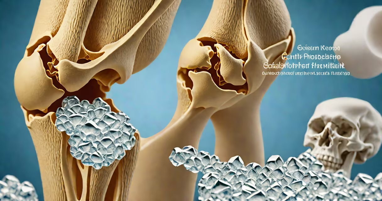
Pseudogout and gout are both forms of crystal arthropathy caused by deposits of microscopic crystals in joints, leading to sudden, painful inflammation. Gout results from monosodium urate crystals formed due to high blood uric acid levels, while pseudogout stems from calcium pyrophosphate dihydrate (CPPD) crystals, which accumulate independently of uric acid. Both conditions mimic severe arthritis but differ in triggers, affected joints, and long-term outcomes.
Both pseudogout and gout develop when crystals form in synovial fluid and deposit in joint tissues. These crystals activate immune cells, triggering a cascade of inflammation that causes pain, swelling, redness, and heat. Attacks typically begin abruptly, often overnight, and last several days to weeks without treatment. The key difference lies in the crystal composition: gout crystals are needle-shaped urate monohydrate, while pseudogout crystals are rhomboid-shaped CPPD crystals.
Diagnosis relies on joint fluid analysis under polarized light microscopy. Urate crystals appear yellow when aligned with the axis of the microscope's polarizer and turn red when rotated 90 degrees. CPPD crystals show a similar birefringent pattern but appear blue-white under the same conditions. This distinction is critical because treatments differ. Misdiagnosing pseudogout as gout leads to ineffective or harmful therapy.
Gout arises from hyperuricemia-elevated serum uric acid levels. When uric acid exceeds solubility limits, it crystallizes into monosodium urate. This process is influenced by diet (high-purine foods like red meat and shellfish), genetics, obesity, kidney dysfunction, and certain medications. Uric acid levels above 6.8 mg/dL increase risk, though not everyone with high levels develops gout.
Pseudogout, conversely, involves calcium pyrophosphate dihydrate (CPPD) crystals. These form when calcium and pyrophosphate concentrations rise in joint fluid. Unlike gout, pseudogout is not linked to uric acid metabolism. Instead, it correlates with aging, joint trauma, hypothyroidism, hemochromatosis, and genetic mutations affecting pyrophosphate metabolism. CPPD deposition is common in older adults-up to 30% of people over 65 show evidence of CPPD crystals in asymptomatic joints.
A 2022 study published in Arthritis & Rheumatology found that among patients with acute knee arthritis, 14% had CPPD crystals, compared to 22% with urate crystals. This highlights that pseudogout is not rare-it occurs nearly as frequently as gout in emergency settings, yet remains underrecognized.
The location of joint attacks differs between the two conditions. Gout most commonly affects the first metatarsophalangeal joint-the big toe-especially in men. This pattern earned gout the nickname "podagra." Other sites include ankles, knees, wrists, and fingers. The classic presentation is a red, hot, swollen toe that wakes a person at night.
Pseudogout preferentially targets large joints. The knee is the most frequent site, followed by the shoulder, wrist, and ankle. A 2021 retrospective review of 1,200 joint aspirations showed that 68% of pseudogout cases involved the knee, versus 76% of gout cases involving the foot or ankle. This difference is clinically useful: a patient with sudden knee swelling and no history of gout should raise suspicion for pseudogout.
| Feature | Gout | Pseudogout |
|---|---|---|
| Crystal Type | Monosodium urate | Calcium pyrophosphate dihydrate (CPPD) |
| Common Joint Sites | Big toe (70%), ankles, knees | Knee (68%), shoulders, wrists |
| Age of Onset | 30-50 years (men), 50+ (women) | 50+ years, peak 60-80 |
| Uric Acid Levels | Elevated in 60-70% of cases | Normal in 90% of cases |
Another key distinction: gout attacks are often recurrent and may progress to chronic tophaceous disease if untreated. Pseudogout episodes tend to be less frequent but can cause long-term joint damage. Repeated CPPD crystal deposition leads to chondrocalcinosis-visible as calcifications on X-rays-and increases risk of osteoarthritis in affected joints.
Many clinicians confuse pseudogout with gout because both present with acute inflammatory arthritis. A 2020 survey of rheologists found that 38% misdiagnosed pseudogout as gout during initial evaluation. This error occurs because both conditions share clinical features: rapid onset, severe pain, joint effusion, and fever in some cases.
Imaging can help differentiate. X-rays reveal characteristic findings: gout shows punched-out erosions in bone near joints, especially in the feet. Pseudogout shows chondrocalcinosis-calcification within cartilage, particularly in the knee's menisci and articular cartilage. However, these signs aren't always visible early in disease. A 2019 study reported that only 45% of pseudogout patients had detectable calcifications on initial X-ray.
One non-obvious fact: pseudogout can mimic septic arthritis. In a 2021 case series from the Mayo Clinic, 12% of patients initially diagnosed with septic arthritis were later found to have CPPD crystals. This underscores the need for joint aspiration in any acute joint infection suspected of being infectious. Treating pseudogout as sepsis with antibiotics risks worsening outcomes.
Treatment diverges sharply due to underlying causes. Gout management focuses on lowering uric acid levels. Allopurinol, febuxostat, and probenecid reduce production or improve excretion of uric acid. These drugs prevent future flares and dissolve existing tophi. Colchicine and NSAIDs treat acute attacks.
Pseudogout treatment does not involve uric acid-lowering agents. NSAIDs, corticosteroids, and colchicine are used acutely, but long-term prevention is not standard. There is no proven therapy to reduce CPPD crystal formation. Some patients benefit from joint aspiration to relieve pressure and remove crystals, but recurrence is common.
I once evaluated a 72-year-old man with a swollen left knee who had been treated for gout with allopurinol for five years. His uric acid was normal, and he had no history of gout flares. After joint aspiration, polarized light microscopy revealed blue-white birefringent crystals-confirming pseudogout. Discontinuing allopurinol resolved his confusion. This case illustrates how treating pseudogout as gout wastes time and exposes patients to unnecessary medication side effects.
Acute joint pain with swelling, redness, and warmth requires prompt assessment. A healthcare provider should perform joint aspiration to identify crystal type. Blood tests for uric acid are useful in gout but often normal in pseudogout. Imaging such as ultrasound or MRI can detect early crystal deposition before symptoms arise.
If you experience sudden joint pain, especially in the knee or big toe, seek evaluation for gout in my toe or similar conditions. Early diagnosis prevents joint damage and guides correct treatment. Misdiagnosis leads to ineffective therapy and prolonged suffering.
Understanding pseudogout vs gout ensures accurate care. While both involve crystal-induced joint inflammation, their causes, patterns, and treatments differ. Recognizing these differences improves outcomes and avoids therapeutic errors.