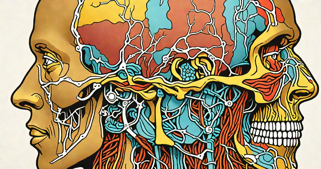
Human face anatomy refers to the structural organization of bones, muscles, nerves, blood vessels, and skin that define the anterior portion of the human skull. This system enables facial expression, sensory perception, and individual identity. Understanding it is essential for medical professionals, artists, and anyone studying human form.
The face functions through integrated systems: skeletal support, muscular movement, neural control, and vascular supply. The underlying structure is formed by eight bones of the skull's facial skeleton, including the maxilla, mandible, zygomatic bones, nasal bones, and frontal bone. These bones provide shape and anchor points for soft tissues.
Facial muscles, such as the orbicularis oculi and zygomaticus major, contract under voluntary and reflex control via the facial nerve (cranial nerve VII). These movements produce expressions like smiling, frowning, and blinking. The trigeminal nerve (cranial nerve V) supplies sensation to the face, detecting touch, temperature, and pain across the forehead, cheeks, and jaw.
For example, the orbicularis oris muscle surrounds the mouth and closes the lips during speech and eating. Its coordinated action with the buccinator muscle helps maintain food between teeth during chewing. Damage to the facial nerve can result in unilateral facial paralysis, as seen in Bell's palsy, where patients lose the ability to smile on one side.
The face consists of four primary tissue layers: skin, subcutaneous fat, muscle, and periosteum (bone membrane). Each layer contributes to function and appearance. Skin varies in thickness across regions-thinner on the eyelids, thicker on the cheeks-and contains sebaceous glands and hair follicles.
Facial muscles are categorized by location and function:
Neurovascular structures follow predictable paths. The external carotid artery branches into the facial artery, which runs along the jawline and supplies blood to the lower face. The facial nerve travels through the parotid gland before dividing into five terminal branches that innervate facial muscles.
Human face anatomy is critical in clinical medicine, especially in plastic surgery, neurology, dentistry, and forensic science. Surgeons rely on precise knowledge to reconstruct faces after trauma or congenital defects. For instance, in cleft lip repair, surgeons must restore the orbicularis oris muscle's continuity to enable normal feeding and speech development.
Dentists use facial anatomy to assess occlusion and plan orthodontic treatments. Misalignment of the maxilla or mandible affects both bite function and facial symmetry. Imaging techniques like CT scans and MRI visualize internal structures such as the paranasal sinuses and temporomandibular joints (TMJ).
Forensic anthropologists analyze facial remains using landmark points-such as the glabella (between the eyebrows) and the gonion (angle of the jaw)-to estimate age, sex, and ancestry. A study published in *The Journal of Forensic Sciences* found that facial measurements correlate strongly with demographic traits, enabling identification from partial remains.
Many assume the nose is primarily cartilage-based, but it includes both cartilage and bone. The nasal septum is made of hyaline cartilage and the perpendicular plate of the ethmoid bone. The upper lateral cartilages form the bridge, while the alar cartilages shape the nostrils. Incorrect assumptions about this structure lead to surgical complications in rhinoplasty.
Another misconception is that facial expressions are universal across cultures. While basic emotions like fear and happiness show similar facial patterns globally, cultural norms modify intensity and context. Research from the University of California, Berkeley, shows that people from collectivist societies often suppress emotional displays, particularly in public settings.
A lesser-known fact: the face contains more than 40 distinct muscles. Most are small and deeply located, such as the levator labii superioris alaeque nasi, which elevates the upper lip and flares the nostrils during sneering. These muscles work in synergy-no single muscle acts alone-making facial motion highly coordinated.
In 2019, a patient presented with drooping of the left side of the face after a viral infection. Physical exam revealed inability to close the left eye, raise the left eyebrow, or smile symmetrically. Neurological testing confirmed facial nerve palsy. MRI showed inflammation around the geniculate ganglion, consistent with Ramsay Hunt syndrome (herpes zoster oticus).
Treatment included antiviral medication (valacyclovir), corticosteroids (prednisone), and eye protection to prevent corneal damage. Recovery took 10 weeks, with gradual return of facial movement. This case illustrates how understanding facial nerve pathways guides diagnosis and intervention.
Human face anatomy is not just a static map of bones and muscles-it is a changing interface between internal physiology and external interaction. From communication to survival, every feature serves a purpose shaped by evolution and function.