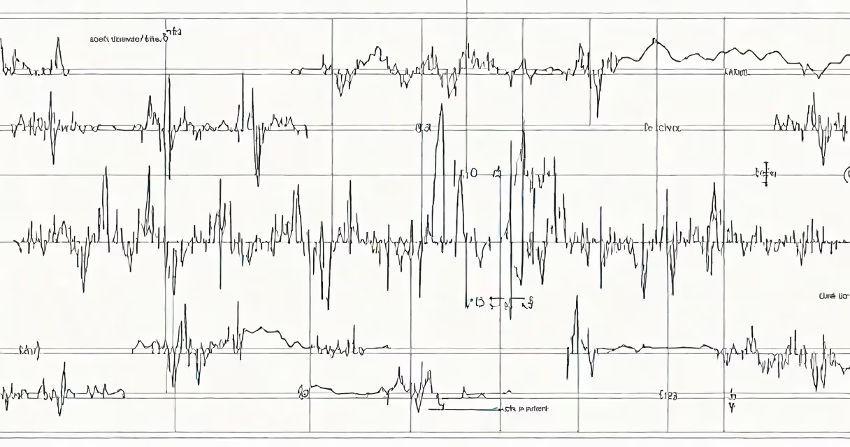
Complete heart block ECG, also known as third-degree atrioventricular (AV) block, occurs when electrical impulses from the atria fail to conduct through the AV node to the ventricles. This results in independent atrial and ventricular rhythms, visible on electrocardiogram as dissociated P waves and QRS complexes.
The condition arises from a complete interruption in conduction between the atria and ventricles. The sinoatrial (SA) node continues to fire normally, producing regular P waves. However, the AV node or His-Purkinje system fails to transmit these signals. As a result, the ventricles generate their own escape rhythm, typically at 30-40 beats per minute, originating from the junction or lower conduction system.
On ECG, complete heart block appears as regular P waves occurring at a consistent rate, while QRS complexes occur independently at a slower, often irregular rate. The PR interval is absent or variable because no relationship exists between P waves and QRS complexes. The QRS morphology depends on the site of the escape pacemaker: narrow if junctional (typically 60-100 ms), wide if ventricular (≥120 ms).
Normal cardiac conduction begins in the SA node, travels through the atria, reaches the AV node, then proceeds via the bundle of His and Purkinje fibers to depolarize the ventricles. In complete heart block, conduction stops at the AV node or below. The ventricles respond with a spontaneous escape rhythm, usually generated by latent pacemaker cells in the AV junction or within the ventricles.
Escape rhythms maintain cardiac output but at a reduced rate. Junctional escape rhythms typically produce a narrow QRS complex and a rate of 40-60 bpm. Ventricular escape rhythms produce wide, bizarre QRS complexes and rates of 20-40 bpm. These rhythms are less efficient and may lead to hemodynamic instability, especially during exertion or stress.
Without intervention, complete heart block can progress to asystole. The risk increases with underlying conditions such as myocardial infarction, cardiomyopathy, or medication toxicity. Immediate treatment is required if symptoms like syncope, hypotension, or chest pain develop.
Complete heart block is not a physical structure but a physiological state defined by ECG criteria. It results from structural or functional disruption in the conduction system. The primary causes include ischemic heart disease, prior cardiac surgery, infiltrative diseases (e.g., sarcoidosis), congenital abnormalities, and drug-induced effects (e.g., beta-blockers, calcium channel blockers).
There are two main types based on the site of block:
| Type | Site of Block | ECG Features | Rate | Prognosis |
|---|---|---|---|---|
| Junctional | AV node or proximal His bundle | Narrow QRS, rate 40-60 bpm | 40-60 bpm | Better hemodynamic tolerance |
| Ventricular | Distal His bundle or Purkinje fibers | Wide QRS, rate 20-40 bpm | 20-40 bpm | Poorer prognosis, higher risk of asystole |
For example, a 72-year-old man with acute inferior wall myocardial infarction developed complete heart block. His ECG showed regular P waves at 70 bpm, QRS complexes at 35 bpm, and no consistent PR relationship. The QRS was wide (140 ms), indicating a ventricular escape rhythm. He required urgent temporary pacing and later permanent pacemaker implantation.
Complete heart block ECG is used in emergency medicine, cardiology, and critical care to diagnose and manage life-threatening arrhythmias. It is a key finding in patients presenting with syncope, dizziness, or fatigue. Early recognition prevents sudden cardiac death.
It is most commonly seen in patients with acute myocardial infarction, particularly those with posterior or inferior wall involvement. The posterior descending artery supplies the AV node in ~90% of people, making it vulnerable during inferior MI. A study published in the Journal of the American College of Cardiology found that complete heart block occurs in 2-3% of acute MI cases and correlates with higher in-hospital mortality.
It also appears in post-cardiac surgery patients, especially after mitral valve repair or coronary artery bypass grafting. In one cohort study, 1.8% of patients developed complete heart block post-op, with a higher incidence in those with pre-existing conduction defects.
Complete heart block is not synonymous with bradycardia. While both involve slow heart rates, bradycardia is a general term for heart rates below 60 bpm. Complete heart block is a specific conduction disorder. A patient can have bradycardia due to sinus node dysfunction without AV block.
Another misconception is that complete heart block always requires a pacemaker. While permanent pacing is indicated in symptomatic cases, asymptomatic patients with stable junctional escape rhythms may be monitored. The 2022 ACC/AHA/HRS guidelines recommend pacing for symptomatic complete heart block, regardless of cause, but allow observation in stable, asymptomatic individuals with adequate ventricular rates.
A non-obvious distinction: complete heart block can mimic artifact or poor lead placement. For example, a wandering baseline or muscle tremor may simulate dissociated P waves. Always verify the rhythm by checking multiple leads. Lead II and V1 are most reliable for detecting P wave morphology and AV dissociation.
I once reviewed a case where a patient with suspected complete heart block had a false-positive reading due to electrode misplacement. After correcting the limb leads, the ECG showed normal AV conduction. This highlights the importance of technical accuracy in ECG interpretation.
Complete heart block ECG is a serious diagnosis requiring prompt evaluation. It is not a standalone event but a sign of underlying pathology. Conditions like Lyme carditis, rheumatic fever, or infiltrative diseases (e.g., amyloidosis) can cause it. A 2021 study in Circulation reported that 15% of complete heart block cases in adults under 50 were due to autoimmune or infectious causes, emphasizing the need for etiological workup.
Understanding complete heart block ECG enables timely intervention. Recognizing the dissociated rhythm, assessing rate and QRS width, and identifying the underlying cause are essential steps in management. Prompt pacing or device therapy improves survival and quality of life.