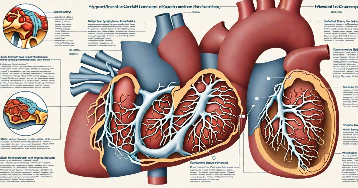
Hypertrophic cardiomyopathy (HCM) is a genetic heart condition characterized by abnormal thickening of the heart muscle, particularly the left ventricle, which can obstruct blood flow and disrupt electrical activity. It affects approximately 1 in 500 individuals worldwide and is a leading cause of sudden cardiac death in young athletes.
The primary issue in HCM is the thickening of the myocardium-the muscular wall of the heart-without an obvious cause such as high blood pressure or valve disease. This thickening often occurs asymmetrically, most commonly near the base of the septum, creating a changing obstruction during systole. The condition results from mutations in genes encoding sarcomere proteins, the contractile units of heart muscle cells.
According to the American Heart Association, HCM accounts for up to 40% of cases of sudden cardiac death in individuals under 35 years old. It is inherited in an autosomal dominant pattern, meaning a single copy of the altered gene from one parent is sufficient to cause the disorder. About 60% of diagnosed cases have a family history of HCM, though new mutations occur in roughly 40% of patients.
HCM arises from mutations in genes such as MYH7, MYBPC3, TNNT2, and TNNI3, which encode components of the cardiac sarcomere. These mutations lead to disorganized myofibrils and inefficient contraction, resulting in increased muscle mass and reduced ventricular compliance. As the heart muscle thickens, the chamber volume decreases, impairing filling during diastole.
When the left ventricular outflow tract narrows due to the thickened septum, turbulent blood flow develops. This creates a gradient measured by echocardiography-typically above 30 mmHg during exercise or stress. The obstruction worsens with increased contractility, such as during physical exertion or anxiety, leading to symptoms like chest pain and dizziness.
Electrical instability is another critical feature. Abnormal tissue architecture disrupts normal conduction pathways, increasing the risk of ventricular arrhythmias. A 2021 study published in *Circulation* found that 15% of HCM patients had sustained ventricular tachycardia, a major contributor to sudden cardiac arrest.
HCM is categorized based on the presence or absence of left ventricular outflow tract obstruction:
| Type | Description | Prevalence | Key Feature |
|---|---|---|---|
| Obstructive HCM | Thickened septum blocks blood flow out of the left ventricle | ~70% | Changing obstruction during systole |
| Non-obstructive HCM | Heart muscle thickens without significant outflow blockage | ~30% | Reduced diastolic filling capacity |
Non-obstructive HCM is often underdiagnosed because symptoms may mimic other conditions like hypertension or heart failure with preserved ejection fraction (HFpEF). However, both forms carry risks of atrial fibrillation and stroke due to impaired relaxation and blood stasis in the left atrium.
A less common variant is apical HCM, where thickening occurs at the tip of the heart rather than the base. This form is more prevalent in Japan and is associated with a distinct pattern on MRI called "reverse T-wave" abnormalities. Apical HCM can mimic coronary artery disease, leading to misdiagnosis if not evaluated with cardiac MRI.
HCM is frequently mistaken for athletic heart syndrome, a benign adaptation seen in trained endurance athletes. Both conditions show increased left ventricular wall thickness, but in athletic heart syndrome, the thickening is symmetric and proportional to body size, with normal diastolic function and no outflow obstruction.
Unlike athlete's heart, HCM shows asymmetric septal hypertrophy, reduced cavity size, and impaired relaxation. A 2019 study in *JACC: Cardiovascular Imaging* showed that 23% of athletes initially diagnosed with HCM were later reclassified as having physiological adaptations after detailed phenotyping.
Another frequent error is assuming HCM only affects men. While early diagnosis often occurs in males due to higher rates of symptom reporting, HCM affects both sexes equally. Women are more likely to present with heart failure symptoms and have a higher mortality rate from HCM-related complications, according to the European Society of Cardiology.
In 2018, a 22-year-old college football player collapsed during practice. Emergency responders found him in ventricular fibrillation. He survived after defibrillation and was later diagnosed with HCM via transthoracic echocardiogram. His echo revealed a 2.5 cm thickened interventricular septum and a 50 mmHg gradient across the left ventricular outflow tract during stress testing.
Genetic testing confirmed a pathogenic variant in the MYBPC3 gene. His father and younger sister also tested positive for the mutation. Despite being asymptomatic prior to collapse, he was implanted with an ICD (implantable cardioverter-defibrillator) due to his high-risk profile. This case illustrates how HCM can remain silent until a life-threatening event occurs.
For individuals with known HCM, regular monitoring is essential. The American College of Cardiology recommends annual echocardiograms and Holter monitoring for those with unexplained syncope or family history of sudden death.
Diagnosis begins with echocardiography, which visualizes wall thickness and assesses outflow obstruction. A wall thickness ≥15 mm in adults (≥13 mm in children) in the absence of other causes is diagnostic. Cardiac MRI provides superior detail of tissue composition and fibrosis, especially useful in non-obstructive cases.
Treatment varies by severity. Mild cases may require lifestyle modification-avoiding intense competitive sports and heavy lifting. Medications such as beta-blockers (e.g., metoprolol) reduce heart rate and improve diastolic filling. For obstructive HCM, septal reduction therapy-either alcohol ablation or surgical myectomy-is effective in relieving symptoms and improving survival.
Patients with high-risk features-including family history of sudden death, unexplained syncope, or abnormal blood pressure response during exercise-should be evaluated for ICD implantation. The 2023 ACC/AHA guidelines recommend ICD placement in patients with a 5-year risk of sudden cardiac death exceeding 5%.
While HCM is incurable, effective management allows many patients to live normal lifespans. Advances in genetic testing and personalized treatment plans continue to improve outcomes.