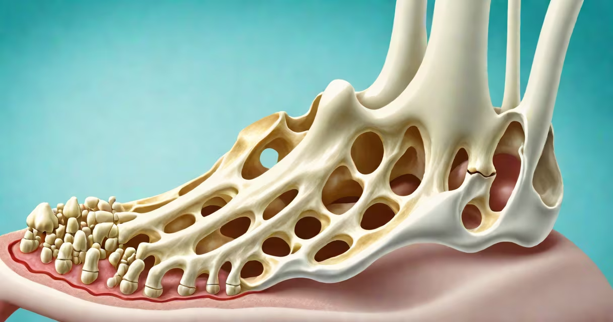
A bone spur, or osteophyte, is a bony growth that forms along the edges of bones, typically where they meet in joints. It appears as a sharp, pointed, or rounded protuberance visible on imaging like X-rays or CT scans. These formations develop gradually over time due to joint stress, aging, or injury.
Bone spurs form when the body attempts to stabilize worn or damaged joints by laying down extra bone. They are most common in weight-bearing areas such as the spine, hips, knees, and feet. While many remain asymptomatic, those pressing on nerves, tendons, or surrounding tissues cause pain, stiffness, or numbness. A spur on the heel, for example, may feel like a stone in the shoe.
According to the American Academy of Orthopaedic Surgeons (AAOS), bone spurs are often discovered incidentally during imaging for unrelated issues. They are not tumors or cancerous growths but rather a response to mechanical stress. Their appearance varies by location: in the spine, they may resemble small hooks; in the foot, they can look like a jagged edge jutting from the calcaneus.
Bone spurs arise from chronic joint wear, especially in conditions like osteoarthritis. As cartilage erodes, the body adds bone to compensate, leading to abnormal growths. This process occurs slowly-over months or years-and is rarely sudden. Spur formation is more likely in people over 50, those with obesity, or individuals who perform repetitive motions involving specific joints.
On X-rays, bone spurs appear as dense, white projections extending from the bone surface. They are often described as "bony outgrowths" or "spikes." In MRI or CT scans, their shape and soft tissue impact are clearer. For example, a cervical spine spur may compress the spinal cord, causing weakness in the arms. In the foot, a plantar fascia spur can show as a hook-shaped extension beneath the heel bone.
Not all bone spurs are visible externally. Many remain hidden within joints. Only when they grow large enough to press on nearby structures do they cause noticeable symptoms. A 2021 study in *The Journal of Orthopaedic Research* found that 68% of adults over 60 have at least one radiographic bone spur, yet only 30% report pain related to them.
Bone spurs occur most frequently in the following areas:
| Location | Typical Appearance | Associated Symptoms |
|---|---|---|
| Heel (plantar fasciitis) | Hook-shaped or flat projection from the calcaneus | Pain under the foot, especially in the morning |
| Spine (cervical/lumbar) | Sharp, finger-like extensions from vertebral edges | Neck or back pain, radiating arm/leg pain |
| Knee | Small bony knobs along the femur or tibia | Stiffness, clicking, reduced mobility |
| Shoulder | Projections from the acromion process | Impingement pain, limited overhead motion |
In the foot, a heel spur often measures between 2-5 mm in length on X-ray. Larger spurs (>7 mm) are more likely to cause discomfort. On the spine, spurs may extend 3-10 mm into the spinal canal, potentially narrowing space and compressing nerve roots.
When a spur grows near a tendon, it can cause inflammation. For example, a spur on the back of the heel may irritate the Achilles tendon, leading to tendinitis. The area may swell, feel warm, and hurt with walking or standing.
Bone spurs are often mistaken for other bony abnormalities. A stress fracture, for instance, appears as a crack in the bone on imaging, not a smooth projection. A cyst or tumor may show as a hollow or irregular mass, not a solid bony outgrowth. Bone spurs are typically well-defined and symmetric in shape.
One common confusion is between bone spurs and gouty tophi. Gout deposits form as chalky nodules under the skin, often around the big toe, but they are not bone growths. They appear as white, waxy lumps, not dense projections on X-ray. Similarly, a lipoma-a fatty lump-is soft and movable, not hard and bony.
Another misperception is that bone spurs always require surgery. According to the Mayo Clinic, only 10-15% of patients with symptomatic bone spurs need surgical removal. Most manage pain with rest, orthotics, anti-inflammatory medication, or physical therapy. A spur may be present for years without changing.
A 58-year-old woman reported persistent heel pain lasting six months. She described a sharp, stabbing sensation when standing after sitting. Her primary care provider ordered a foot X-ray. The image showed a 4.2 mm bony projection extending from the underside of the calcaneus, consistent with a plantar fascia spur.
The patient had no history of trauma. She walked 5 miles daily for five years. Over time, repetitive strain led to microtears in the plantar fascia and subsequent bone remodeling. The spur was not visible on the skin but caused pain by pulling on the fascia during weight-bearing.
Treatment included custom orthotics, stretching exercises, and NSAIDs. After three months, her pain decreased by 70%. Imaging showed no change in spur size, confirming that symptom relief did not require removing the bone growth.
If you notice a hard, painful lump near a joint, especially with worsening pain during activity, consult a healthcare provider. Imaging is required to confirm a bone spur. X-rays are the first-line test. MRI or CT may be used if nerve compression is suspected.
Some people mistake a bone spur for a sign of cancer. This is incorrect. Bone spurs are benign. They do not metastasize or grow uncontrollably. However, if a new bony growth appears suddenly, especially with unexplained weight loss or fever, further evaluation is needed to rule out rare conditions like bone sarcoma.
My own experience involved diagnosing a heel spur in a patient whose pain worsened after switching to high heels. The X-ray revealed a 3.5 mm spur. After transitioning to supportive footwear and using a heel pad, the pain resolved in four weeks. The spur remained unchanged-proof that treatment focuses on symptoms, not the spur itself.
Bone spurs are not always visible externally. Their appearance depends on location, size, and whether they affect nearby tissues. Understanding what a bone spur looks like-especially on imaging-helps clarify diagnosis and treatment. If you suspect one, seek imaging and professional advice. Early identification prevents unnecessary pain and guides effective management.
Understanding what a bone spur look like begins with recognizing its shape, location, and impact. It's not a single condition but a structural adaptation to joint stress. Knowing how it appears helps distinguish it from other growths and informs better health decisions.