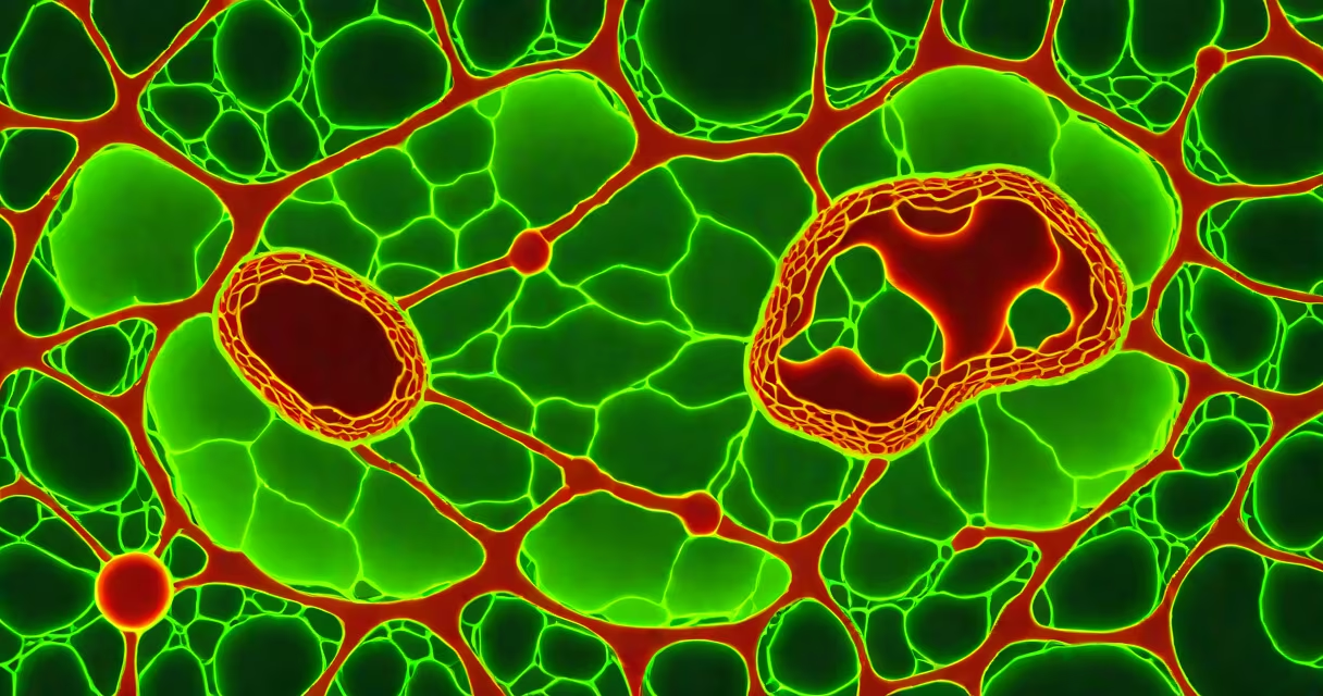
the powerhouse of the cell is a nickname for mitochondria, organelles that generate most of a eukaryotic cell's adenosine triphosphate (ATP) through oxidative phosphorylation. These structures convert chemical energy from food into usable energy for cellular processes.
Mitochondria are double-membrane-bound structures. The outer membrane is smooth, while the inner membrane folds into cristae-increasing surface area for electron transport chain proteins. The space between membranes is the intermembrane space; the innermost region is the matrix, where enzymes for the Krebs cycle reside.
Each human cell contains hundreds to thousands of mitochondria, depending on metabolic demand. Muscle cells, neurons, and liver cells have high densities due to their energy needs. Mitochondria are semi-autonomous-they carry their own DNA (mtDNA), which encodes 13 proteins involved in ATP synthesis, 22 tRNAs, and 2 rRNAs.
ATP production begins when glucose and fatty acids enter the mitochondrial matrix. Pyruvate from glycolysis is transported in and converted to acetyl-CoA, which enters the Krebs cycle. This generates electron carriers-NADH and FADH₂-that shuttle electrons to the inner membrane.
Electrons move through four protein complexes (I-IV) embedded in the inner membrane. As electrons pass through, protons (H⁺) are pumped from the matrix into the intermembrane space, creating a proton gradient. This gradient drives ATP synthase, a rotary enzyme that synthesizes ATP from ADP and inorganic phosphate.
For every 10 protons flowing through ATP synthase, approximately 3 ATP molecules are produced. This process is highly efficient: one glucose molecule can yield up to 36 ATP molecules when fully oxidized in mitochondria, compared to just 2 from glycolysis alone.
While all mitochondria share core functions, their morphology and distribution vary by tissue. Cardiac muscle cells contain densely packed mitochondria arranged in rows along myofibrils, supporting constant contraction. Neurons show mitochondrial clustering near synapses, where energy demands spike during neurotransmission.
Some cells exhibit specialized mitochondrial forms. Brown adipose tissue contains multilocular mitochondria rich in uncoupling protein 1 (UCP1), which dissipates the proton gradient as heat-a process called thermogenesis. This adaptation helps maintain body temperature in newborns and hibernating animals.
| Cell Type | Number of Mitochondria per Cell | Specialized Feature |
|---|---|---|
| Liver hepatocyte | 1,000-2,000 | High enzyme activity for detoxification and gluconeogenesis |
| Cardiomyocyte | 5,000+ | Aligned in sarcomeres; supports sustained contraction |
| Neuron (cerebral cortex) | 100-500 | Localized near axon hillocks and synaptic terminals |
| Brown fat cell | 10,000+ | Contains UCP1 for non-shivering thermogenesis |
People often assume mitochondria are solely responsible for energy production. While they are the primary site of ATP synthesis, other pathways contribute. Glycolysis in the cytoplasm produces 2 ATP per glucose molecule. Some cells, like red blood cells, lack mitochondria entirely and rely exclusively on anaerobic glycolysis.
A common error is equating mitochondrial dysfunction with aging. While mtDNA mutations accumulate with age and correlate with reduced ATP output, they are not the sole cause of aging. The relationship is complex-some studies suggest mitochondrial dynamics (fusion/fission) play a larger role in cellular decline than mutation load alone.
Another misconception: mitochondria evolved from bacteria. This is correct-endosymbiotic theory posits that mitochondria originated from alphaproteobacteria engulfed by ancestral eukaryotes. However, they are not free-living organisms. They cannot survive outside host cells and have lost most ancestral genes, transferring essential ones to the nuclear genome.
Defects in mitochondrial function underlie over 50 known genetic disorders. Leigh syndrome, caused by mutations in mitochondrial complex I subunits, leads to progressive neurological degeneration in infants. Symptoms include developmental delay, seizures, and respiratory failure. Diagnosis often involves measuring lactate levels and analyzing mtDNA sequencing.
In cancer biology, tumor cells frequently exhibit altered mitochondrial metabolism. The Warburg effect describes how many cancer cells favor glycolysis even in oxygen-rich environments. This shift may support rapid biomass production rather than efficient ATP generation. Targeting mitochondrial metabolism is now a focus in experimental oncology.
I once observed a live-cell imaging experiment where fluorescently labeled mitochondria were tracked in fibroblasts. When the cells were deprived of oxygen, the mitochondria fragmented rapidly and moved toward the nucleus-a response linked to apoptosis initiation. This visual confirmed the role of mitochondria not just in energy supply, but in cell death signaling.
While the term "the powerhouse of the cell" accurately describes mitochondria's central role in ATP synthesis, it's misleading to imply they are the only source. Cells also produce ATP via substrate-level phosphorylation in glycolysis and the Krebs cycle. Mitochondria amplify this output through oxidative phosphorylation, but they are not the origin of all energy conversion.
For example, in yeast cells grown under anaerobic conditions, ATP is generated solely through glycolysis. The mitochondria remain intact but inactive. This shows that mitochondria are not always operational-even when present.
Understanding the full context of cellular energy requires recognizing that mitochondria are changing systems, not static generators. Their behavior changes with nutrient availability, oxygen levels, and cellular stress signals.