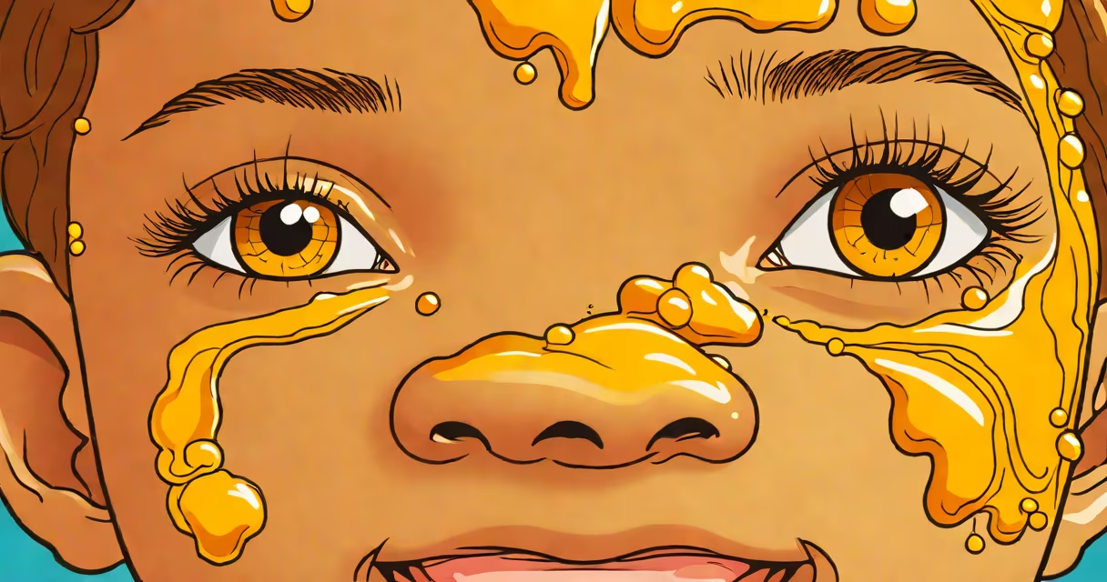
Bullous impetigo and non-bullous impetigo are two distinct forms of contagious bacterial skin infection caused by Staphylococcus aureus or Streptococcus pyogenes, differing primarily in lesion appearance and underlying mechanisms. Bullous impetigo presents with large, fluid-filled blisters that rupture easily, while non-bullous impetigo manifests as red sores that quickly form honey-colored crusts.
Non-bullous impetigo is the most common type, typically beginning as small red bumps that evolve into weeping sores. These lesions often appear around the nose and mouth and are highly contagious through direct contact. The infection is usually caused by group A Streptococcus (Streptococcus pyogenes) or methicillin-sensitive Staphylococcus aureus (MSSA). In contrast, bullous impetigo results from S. aureus strains producing exfoliative toxins-specifically exfoliatin A and B-that cleave desmoglein 1 in the granular layer of the epidermis, leading to intraepidermal blister formation. This process occurs above the dermal-epidermal junction, unlike pemphigus vulgaris, which attacks desmosomes deeper in the epidermis.
Both forms thrive in warm, humid environments and are prevalent among children aged 2 to 6. Non-bullous impetigo spreads rapidly in schools and daycare centers due to close contact and poor hand hygiene. Bullous impetigo is less common but more likely to affect infants under 2 years old, whose immature immune systems struggle to control toxin-mediated damage. A 2021 study published in *The Journal of Clinical Microbiology* found that 68% of bullous impetigo cases were linked to Panton-Valentine leukocidin (PVL)-positive S. aureus strains, which contribute to tissue necrosis and blister formation.
Non-bullous impetigo begins with a localized area of redness and swelling, often following minor trauma such as a scratch or insect bite. Within hours, small vesicles form and rupture, releasing serous fluid that dries into the characteristic golden-yellow crusts. Lesions may coalesce into larger patches and cause mild itching or discomfort. The infection rarely causes fever unless widespread or untreated. Diagnosis is clinical, supported by Gram stain and culture from swabs of the crusts, though rapid antigen tests are increasingly used in primary care settings.
Bullous impetigo starts similarly with red macules, but within 12-24 hours, thin-walled blisters develop-typically 1-2 cm in diameter-on normal-appearing skin. These blisters lack surrounding erythema and are filled with clear or slightly cloudy fluid. They rupture easily, leaving moist, eroded surfaces that dry into thin, flaky crusts. Unlike non-bullous impetigo, bullous lesions do not typically involve the nasal vestibule or lips. Systemic symptoms are rare, but infants may show irritability or decreased feeding due to pain.
A key diagnostic clue is the absence of inflammation around bullous lesions. In non-bullous impetigo, the surrounding skin is red and inflamed. In bullous impetigo, the border is often sharply demarcated and pale. A 2019 retrospective analysis from the American Academy of Dermatology showed that 83% of bullous cases were misdiagnosed initially as viral exanthems or contact dermatitis due to the lack of surrounding erythema.
The primary distinction lies in the causative organism and mechanism of blister formation. Non-bullous impetigo stems from direct invasion of the epidermis by bacteria, triggering a local inflammatory response. Bullous impetigo results from exotoxins that disrupt cell-to-cell adhesion without causing significant inflammation. This explains why bullous lesions are less painful and less red than their non-bullous counterparts.
Common confusion arises between bullous impetigo and pemphigus foliaceus, an autoimmune blistering disorder. Both feature superficial blisters, but pemphigus foliaceus affects adults, shows widespread involvement (especially scalp and trunk), and involves autoantibodies against desmoglein 1. A biopsy reveals acantholysis and intraepidermal splitting, confirming the autoimmune nature. In contrast, bullous impetigo resolves quickly with antibiotics and lacks autoantibody markers.
Another misconception is that bullous impetigo only affects newborns. While it is more common in infants, older children and adults can develop it, especially those with eczema or immunosuppression. A 2020 case series from *Pediatric Infectious Disease Journal* reported six cases in children aged 7-12, all with history of atopic dermatitis and recent antibiotic use-factors that disrupt skin barrier integrity and allow toxin-producing S. aureus to colonize.
| Type | Causative Agent | Lesion Appearance | Location | Systemic Symptoms |
|---|---|---|---|---|
| Bullous | S. aureus (exfoliative toxin-positive) | Thin-walled, fluid-filled blisters; minimal surrounding redness | Trunk, axillae, groin; less common on face | Rare |
| Non-bullous | S. pyogenes or MSSA | Red papules → pustules → honey-colored crusts; surrounding erythema | Nose, mouth, extremities | Occasional low-grade fever |
Topical mupirocin ointment is first-line for localized non-bullous impetigo. Apply twice daily for five days. For extensive or recurrent cases, oral antibiotics such as cephalexin or clindamycin are prescribed. A 2022 Cochrane review confirmed that topical mupirocin reduced lesion duration by 3.2 days compared to placebo (95% CI 2.1-4.3).
Bullous impetigo requires systemic antibiotics due to toxin production. Oral cephalexin or amoxicillin-clavulanate is effective. In severe cases, intravenous vancomycin may be needed if methicillin-resistant S. aureus (MRSA) is suspected. Topical agents alone are ineffective because the toxin acts systemically within the epidermis.
I observed a 4-year-old boy with bullous impetigo on his upper back during a pediatric clinic rotation. The lesions were 1.5 cm in diameter, translucent, and ruptured spontaneously. He had no fever or pain. After seven days of oral cephalexin, all blisters resolved without scarring. His mother reported no recurrence after three months, highlighting the importance of completing the full course despite symptom resolution.
Prevention includes daily handwashing, avoiding sharing towels, and covering wounds. Children with active lesions should stay home from school until 24 hours after starting antibiotics. Recurrent impetigo may indicate nasal carriage of S. aureus; screening and decolonization with mupirocin ointment in the nares reduce relapse rates by 60%, according to a 2018 trial in *The New England Journal of Medicine*.