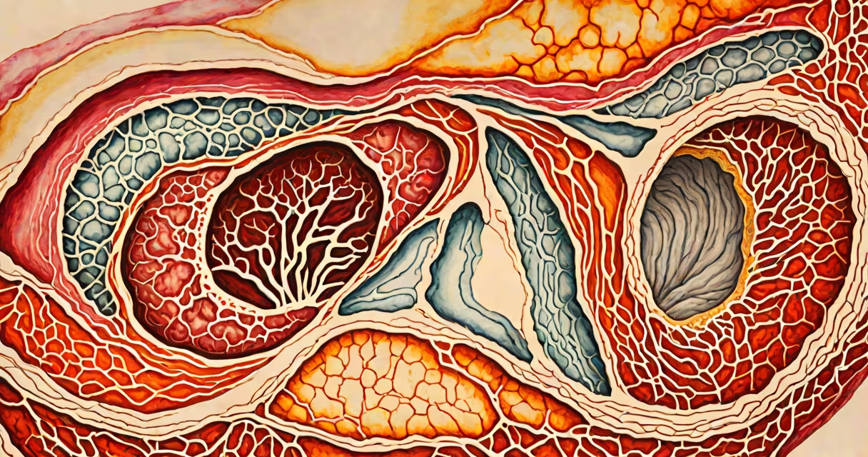
The uterine lining, or endometrium, is a changing layer of tissue composed primarily of epithelial cells, stromal fibroblasts, and a rich network of blood vessels. It thickens under hormonal influence during the menstrual cycle and sheds during menstruation if pregnancy does not occur.
The endometrium is not a single substance but a living tissue system that changes daily. It consists of three functional layers: the stratum basalis (deepest), the stratum functionalis (middle and shed), and the stratum compactum (dense upper layer). These layers are made of connective tissue, glands, and capillaries that respond to fluctuating levels of estrogen and progesterone.
Estrogen, produced by the ovaries during the follicular phase, stimulates proliferation of endometrial cells. Progesterone, released after ovulation, prepares the lining for potential implantation by increasing gland secretion and vascular density. If fertilization fails, hormone levels drop sharply, triggering shedding of the functionalis layer-menstruation.
The endometrium contains several distinct cell types working together. Epithelial cells form the surface barrier and secrete mucus and growth factors. Stromal cells provide structural support and differentiate into decidual cells during early pregnancy. Immune cells such as macrophages and natural killer cells regulate inflammation and tissue remodeling.
Blood vessels in the endometrium are highly responsive. Spiral arterioles remodel dramatically each cycle, becoming narrow and coiled. When progesterone drops, these vessels constrict and rupture, causing bleeding. This process is regulated by angiogenic proteins like VEGF and anti-angiogenic factors such as thrombospondin-1.
A 2021 study published in *Human Reproduction* found that women with recurrent miscarriages often have abnormal endometrial receptivity markers, including altered integrin expression and disrupted HOXA10 gene activity. This shows that the lining's molecular makeup directly affects fertility outcomes.
The endometrium undergoes four phases tied to the menstrual cycle: menstruation, follicular, ovulatory, and luteal. During menstruation, the functionalis layer detaches and exits through the vagina. This phase lasts 3-7 days and involves breakdown of tissue and blood vessels.
In the follicular phase (days 6-14), rising estrogen drives rapid regeneration of the endometrium. Glands elongate, and stroma becomes edematous. By day 14, the endometrium reaches peak thickness-typically 8-14 mm in fertile women.
After ovulation, progesterone dominates. The endometrium shifts from proliferative to secretory. Glands begin secreting glycogen-rich fluid, and stroma becomes more cellular. This secretory phase peaks around day 21-23, creating an optimal environment for embryo implantation.
| Phase | Hormone Dominance | Endometrial Changes | Duration |
|---|---|---|---|
| Menstrual | Low estrogen & progesterone | Tissue shedding, bleeding | 3-7 days |
| Follicular | Estrogen | Proliferation, gland formation | Days 6-14 |
| Ovulatory | Peak estrogen | Surface epithelium thins, mucus increases | Day 14 |
| Luteal | Progesterone | Secretion, vascular remodeling | Days 21-28 |
Abnormal endometrial development is linked to infertility, heavy menstrual bleeding, and endometriosis. Endometriosis occurs when endometrial-like tissue grows outside the uterus, causing pain and scarring. Despite similar histology, this tissue responds differently to hormones and lacks proper shedding mechanisms.
A common misconception is that the uterine lining is simply "mucus." It is not. While it produces mucus, its primary function is to support embryo implantation and act as a barrier against pathogens. The lining also prevents adhesion between the uterine walls during menstruation.
Another misunderstanding is that the endometrium regenerates from stem cells in the basal layer. Research confirms that endometrial progenitor cells reside in the endometrial crypts and are activated during repair. A 2019 study in *Nature Cell Biology* identified a population of Lgr5+ stem cells responsible for tissue renewal after menstruation.
In assisted reproduction, doctors perform endometrial biopsies to assess receptivity. A small sample is taken from the uterine lining during the window of implantation (around day 20 of a 28-day cycle). The tissue is analyzed for gene expression patterns related to implantation success.
For example, a patient undergoing IVF had a biopsy showing low expression of the gene *HOXA10*, which regulates endometrial differentiation. Her doctor prescribed progesterone supplementation and delayed embryo transfer. After treatment, her endometrium thickened to 10 mm, and she achieved a successful pregnancy.
This case illustrates that the uterine lining is not passive-it actively communicates with embryos via cytokines and adhesion molecules. Its composition determines whether pregnancy can proceed.
The endometrium is not the same as the endocervix. The cervical mucus changes during the cycle too, but it serves a different purpose-facilitating sperm transport. The endometrium focuses on implantation and nutrient exchange.
Also, the lining does not regenerate uniformly. The functionalis layer sheds completely, but the stratum basalis remains intact. This residual layer acts as a reservoir for regrowth. Without it, endometrial regeneration would fail-leading to conditions like Asherman's syndrome, where scar tissue forms after trauma.
One non-obvious fact: the endometrium can metabolize glucose and lactate independently of the bloodstream. A 2022 study in *Cell Metabolism* showed that endometrial stromal cells use aerobic glycolysis during the secretory phase, providing energy for tissue remodeling.
I once observed a frozen-thawed endometrial biopsy slide under a microscope during a research rotation. The stromal cells were visibly swollen and filled with glycogen granules-evidence of active preparation for implantation. That moment confirmed how changing and metabolically active the lining truly is.
Understanding what the uterine lining is made of clarifies why hormonal imbalances, infections, or surgical trauma can disrupt fertility. It is not inert tissue-it is a responsive organ shaped by hormones, immune signals, and metabolic demands.