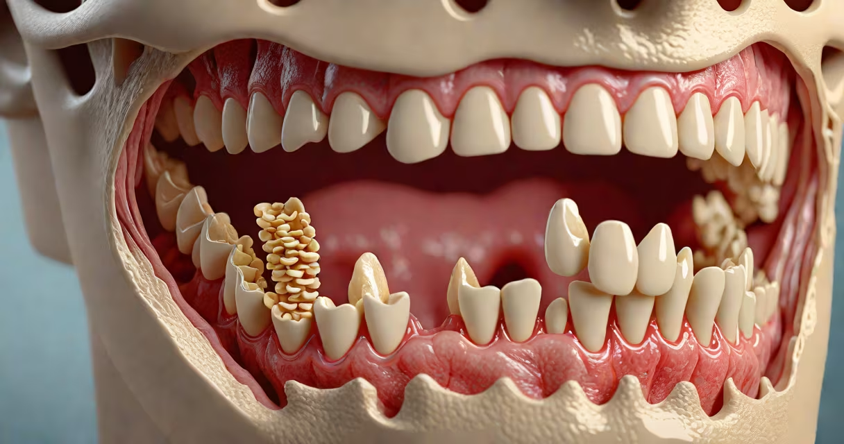
The hard palate is the bony, rigid anterior portion of the roof of the mouth formed by the maxillary and palatine bones. It separates the oral cavity from the nasal cavity and provides structural support for the tongue during speech and mastication.
Located at the front of the oral roof, the hard palate is composed of two distinct bones: the horizontal plates of the maxilla and the palatine bones. These fuse during early development to form a solid, immovable platform. The surface is covered by a thick layer of stratified squamous epithelium, which resists abrasion from food and chewing forces. This tissue is rich in blood vessels and nerves, particularly the greater palatine nerve, which supplies sensation.
Developmentally, the hard palate forms between the sixth and ninth weeks of gestation. Failure of the palatal shelves to fuse properly results in cleft palate, a congenital condition affecting approximately 1 in 1,600 live births globally, according to the Centers for Disease Control and Prevention (CDC).
The hard palate consists of two primary bony components: the maxillary bones and the palatine bones. The maxillary bones contribute the larger anterior portion, while the palatine bones form the posterior segment. The junction between these bones is marked by the palatine raphe-a visible ridge of connective tissue running along the midline.
The mucosal lining of the hard palate contains numerous minor salivary glands that secrete mucus to lubricate the oral environment. These glands are essential for maintaining oral hygiene and facilitating speech articulation. The presence of keratinized epithelium protects against mechanical trauma from food particles and dental instruments.
A key anatomical landmark is the incisive canal, located near the center of the hard palate behind the upper central incisors. This small passage allows the passage of the nasopalatine nerve and blood vessels into the nasal cavity. Clinically, this area is significant during local anesthesia procedures for dental work.
| Component | Bone(s) Involved | Location | Function |
|---|---|---|---|
| Anterior hard palate | Maxillary horizontal plate | Front half, under the nose | Supports tongue, aids in speech |
| Posterior hard palate | Palatine bone | Back half, near uvula | Seals nasal cavity from mouth |
| Palatine raphe | Fibrous tissue | Midline ridge | Visible fusion line from embryonic development |
The hard palate plays a critical role in speech production. During consonant articulation-especially alveolar sounds like "t," "d," and "n"-the tip of the tongue contacts the hard palate to create obstruction and airflow modulation. Without this contact point, speech clarity diminishes.
In digestion, the hard palate stabilizes the tongue during mastication. As the tongue moves food toward the pharynx, the firm surface prevents the tongue from collapsing inward. This enables efficient bolus formation and safe swallowing. Individuals with cleft palate often require surgical repair and speech therapy to compensate for disrupted function.
A study published in *The Journal of Craniofacial Surgery* found that patients with repaired cleft palates had 85% improved articulation accuracy after multidisciplinary intervention, including speech pathology and orthodontic alignment.
Many confuse the hard palate with the soft palate, which lies posterior and is composed of muscle and connective tissue. The soft palate elevates during swallowing to close off the nasopharynx, preventing food from entering the nasal cavity. The hard palate remains static and bony throughout life.
Another misconception is that the hard palate is purely structural. In reality, it contributes to sensory feedback during eating. Nerve endings in the mucosa detect texture, temperature, and pressure, influencing taste perception and satiety signals.
Oral cancer can develop on the hard palate, particularly in smokers and users of smokeless tobacco. A lesion lasting more than two weeks with pain, bleeding, or ulceration requires biopsy. The American Cancer Society reports that 3% of all oral cancers originate in the hard palate region.
I once observed a patient undergoing a dental implant procedure where the surgeon used a custom surgical guide based on CT scans to avoid damaging the maxillary sinus through the hard palate. The guide ensured precise placement of the implant in the posterior region, avoiding the thin cortical bone and minimizing risk of infection or sinus perforation.
This case highlighted how understanding hard palate anatomy is not just academic-it directly impacts surgical outcomes. Even minor errors in navigation can lead to complications such as oroantral fistulas, requiring additional corrective surgery.
The hard palate connects to several important structures. Superiorly, it borders the nasal cavity via the ethmoid bone and sphenoid sinuses. Inferiorly, it supports the maxillary teeth and forms the floor of the nasal cavity. The palatine arteries supply blood, while the greater palatine nerve provides sensory innervation.
During a routine dental exam, dentists palpate the hard palate to assess for asymmetry, tenderness, or abnormal growths. Any deviation from symmetry may indicate underlying pathology, such as a torus palatinus-a benign bony growth that occurs in 3-4% of the population, most commonly in adults over 40.
The hard palate is not merely a barrier-it is a functional interface between breathing, speaking, and eating. Its stability enables complex behaviors we take for granted daily. Understanding its anatomy clarifies both normal physiology and the basis for many clinical interventions.
For those seeking deeper insight into cranial anatomy, exploring the relationship between the hard palate and the temporomandibular joint offers further context on jaw mechanics and facial development.