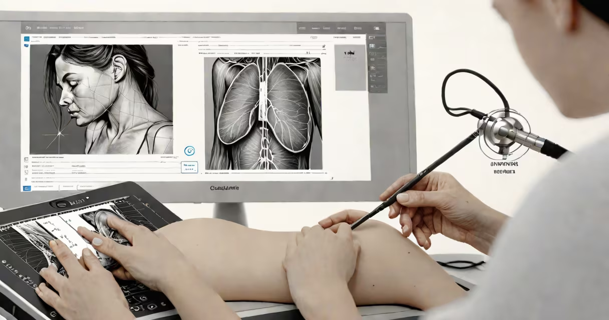
Ultrasound of the breast uses high-frequency sound waves to create real-time images of breast tissue, revealing structures such as cysts, solid masses, and blood flow patterns. It differentiates between fluid-filled cysts and solid tumors, helping assess whether a finding requires biopsy. This non-invasive tool complements mammography, especially in dense breast tissue.
Unlike X-ray-based methods, ultrasound does not use ionizing radiation. Instead, it relies on echoes from sound waves bouncing off internal tissues. A transducer emits pulses into the breast and captures returning signals. These signals are processed into cross-sectional images displayed on a monitor within seconds. The procedure is painless and typically takes 15-30 minutes.
Ultrasound is particularly effective in evaluating palpable lumps, unclear mammogram results, or asymmetries in breast density. It is often used during pregnancy and in younger women whose breasts are denser, where mammograms have lower sensitivity. Radiologists interpret findings based on shape, margins, echogenicity, and Doppler signal for vascularity.
The process begins with the patient lying supine or slightly tilted. A water-based gel is applied to the skin to reduce air gaps and improve sound transmission. The sonographer moves the transducer slowly across the breast, scanning each quadrant systematically. Images are captured in multiple planes-longitudinal and transverse-to assess lesion orientation and depth.
Key features assessed include size, shape, margin regularity, internal texture (homogeneous vs. heterogeneous), and posterior shadowing. For example, a round mass with smooth edges and no shadowing may suggest a benign fibroadenoma. In contrast, a irregular, hypoechoic (dark) mass with microlobulated margins raises concern for malignancy.
Doppler ultrasound adds functional insight by detecting blood flow. Malignant tumors often develop abnormal, high-velocity vessels. A study published in *Radiology* found that Doppler features improved specificity in distinguishing cancer from benign lesions by 18% compared to grayscale alone.
Ultrasound identifies several breast conditions based on visual and acoustic properties:
| Condition | Appearance on Ultrasound | Common Location |
|---|---|---|
| Cyst | Round, anechoic (black), thin-walled, with posterior enhancement | Upper outer quadrant, often solitary |
| Fibroadenoma | Well-defined, oval, homogeneous echo pattern, mobile | Mid-outer breast, common in women under 35 |
| Intraductal papilloma | Small, solid mass near nipple, may show color Doppler signal | Nipple-areolar complex |
| Carcinoma (invasive ductal) | Irregular shape, spiculated margins, hypoechoic, posterior shadowing | Variable, often in upper outer quadrant |
For instance, a 42-year-old woman with a 1.2 cm lump in her left breast underwent ultrasound after a suspicious mammogram. The lesion was irregular, had indistinct borders, and showed increased vascularity on Doppler. Biopsy confirmed invasive ductal carcinoma. Without ultrasound, the subtle features might have been missed on mammography alone.
Ultrasound also guides needle placement during biopsy or drainage. In one clinical trial, ultrasound-guided core biopsy achieved 96% accuracy in diagnosing breast cancer, according to the American College of Radiology.
Many patients assume ultrasound replaces mammography. That is incorrect. Ultrasound detects different types of abnormalities and cannot visualize microcalcifications-key early signs of ductal carcinoma in situ (DCIS). Mammography remains the gold standard for population screening.
Another misconception is that all solid masses are cancerous. In reality, most solid breast masses detected by ultrasound are benign. Fibroadenomas account for nearly 60% of solid masses in women under 40, according to the National Cancer Institute.
Ultrasound is operator-dependent. Image quality varies based on the sonographer's skill and equipment resolution. Low-frequency probes may miss small lesions. High-resolution linear probes (10-15 MHz) are preferred for breast imaging.
A critical but often overlooked point: ultrasound cannot reliably stage breast cancer. It shows local extent but not lymph node involvement or distant metastasis. Staging still requires MRI, CT, or PET scans.
Ultrasound is routinely ordered when a mammogram reveals a suspicious area, especially in women with dense breasts. The American College of Radiology recommends ultrasound for women with heterogeneously or extremely dense breasts who have a negative mammogram but a palpable lump.
It is also used to evaluate nipple discharge. If bloody or unilateral, ultrasound can detect intraductal lesions. In one study, ultrasound identified 83% of papillomas causing discharge, reducing unnecessary surgical exploration.
I once observed a case where a 38-year-old woman presented with a firm, mobile lump in her right breast. Mammography showed no calcifications or architectural distortion. Ultrasound revealed a well-circumscribed, oval mass with posterior enhancement-classic for a fibroadenoma. She avoided biopsy and was monitored annually. After two years, the mass remained stable.
For women undergoing breast augmentation, ultrasound helps assess implant integrity and detect capsular contracture. It is also used post-lumpectomy to check for residual disease or recurrence.
Findings are categorized using BI-RADS (Breast Imaging Reporting and Data System) scores. A score of 3 indicates probably benign, with follow-up in 6 months. Score 4 suggests intermediate suspicion, requiring biopsy. Score 5 indicates high likelihood of cancer-biopsy is recommended.
If ultrasound shows a complex cyst (part solid, part fluid), the radiologist may recommend aspiration or monitoring. A 2021 meta-analysis in *European Radiology* found that complex cysts with solid components had a 12% cancer risk, warranting close follow-up.
For those with a family history of breast cancer, ultrasound may be added to annual screening. However, routine screening in average-risk women without symptoms is not recommended due to false positives and overdiagnosis.
Ultrasound of the breast shows soft-tissue details invisible to mammography. It identifies cysts, evaluates solid masses, guides biopsies, and monitors treatment response. While not a standalone screening tool, it is essential in refining diagnoses and avoiding unnecessary procedures.