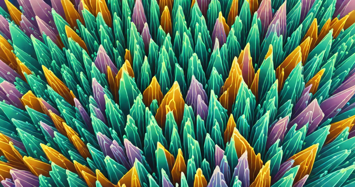
Uric acid crystals form when excess uric acid in the bloodstream exceeds its solubility threshold, precipitating into sharp, needle-shaped deposits in joints, tissues, or kidneys. This crystallization drives inflammation and tissue damage, most commonly manifesting as gout or kidney stones.
Uric acid is a waste product generated during the breakdown of purines-nitrogen-containing compounds found in DNA and certain foods. Normally, the kidneys filter out excess uric acid, excreting it through urine. When production overwhelms elimination, serum levels rise, leading to hyperuricemia. Once concentrations surpass ~6.8 mg/dL, supersaturation occurs, enabling nucleation and growth of monosodium urate (MSU) crystals.
These crystals trigger immune responses via activation of the NLRP3 inflammasome, releasing interleukin-1β and recruiting neutrophils. This cascade results in acute joint pain, swelling, redness-hallmarks of gout flares. Crystals can also accumulate in renal tubules, causing nephrolithiasis or chronic kidney disease over time.
Crystallization begins when uric acid concentration exceeds saturation levels in extracellular fluid. Solubility depends on temperature, pH, and ion concentration. At physiological pH (7.4), uric acid exists primarily as urate anions, which bind sodium to form MSU crystals. These crystals grow slowly but persist due to their low solubility and resistance to enzymatic degradation.
Once formed, crystals act as foreign bodies. Macrophages attempt phagocytosis but fail, releasing pro-inflammatory cytokines. Neutrophils swarm in, degranulating and producing reactive oxygen species. This inflammatory storm explains the intense pain and rapid onset of gout attacks-often within hours.
A 2021 study published in *Nature Reviews Rheumatology* confirmed that MSU crystals induce pyroptosis, a form of programmed cell death that amplifies inflammation beyond initial immune cell activation. This mechanism underscores why even small crystal burdens can cause severe clinical symptoms.
Excess uric acid arises from either increased production or decreased excretion. About 80-90% of cases stem from impaired renal excretion; the remainder result from overproduction, often due to enzyme defects or high-purine intake.
| Category | Specific Cause | Prevalence/Impact |
|---|---|---|
| Increased Production | Overactive xanthine oxidase enzyme | Accounts for ~10% of gout cases; seen in Lesch-Nyhan syndrome |
| Decreased Excretion | Reduced function of URAT1 transporter in proximal tubules | Genetic variants affect ~30% of hyperuricemic individuals |
| Dietary Influence | High intake of red meat, shellfish, alcohol (especially beer) | Beer contains purines and ethanol, both increasing uric acid synthesis |
| Comorbid Conditions | Hypertension, insulin resistance, metabolic syndrome | Each independently reduces renal clearance by 15-25% |
Alcohol consumption is particularly potent. Ethanol increases uric acid levels through multiple pathways: inhibiting renal excretion, accelerating ATP breakdown (producing more purines), and reducing uricase activity in humans (unlike many mammals).
Non-obvious fact: Even low-dose diuretics like hydrochlorothiazide reduce uric acid excretion by competing with urate transporters in the proximal tubule. A 2019 meta-analysis in *The Lancet* showed patients on thiazides had a 40% higher risk of developing gout compared to non-users.
Many assume gout is purely a "rich man's disease" linked to overindulgence. While diet plays a role, genetics and comorbidities are stronger predictors. A 2022 study in *Arthritis & Rheumatology* found that 60% of gout patients had no identifiable dietary triggers-instead, they carried variants in SLC2A9 and ABCG2 genes affecting urate transport.
Another misconception: All crystals are harmful. In reality, asymptomatic hyperuricemia affects ~20% of adults globally, yet only 10-20% develop symptomatic gout. The presence of crystals doesn't guarantee inflammation-host factors like immune response thresholds determine clinical outcomes.
Also, not all joint pain from crystals is sudden. Chronic tophaceous gout involves slow deposition of large crystal aggregates (tophi) in cartilage, tendons, or earlobes. These may remain silent for years until trauma or infection triggers flare-ups.
A 52-year-old male presented with sudden left big toe pain lasting 48 hours. He reported drinking six beers weekly and eating red meat twice weekly. Blood tests revealed serum uric acid of 9.2 mg/dL. Synovial fluid aspiration showed negatively birefringent, needle-shaped crystals under polarized light-diagnostic for MSU crystals.
He was diagnosed with acute gout. After treatment with colchicine and NSAIDs, his pain resolved in 72 hours. His uric acid remained elevated despite lifestyle changes. Genetic testing revealed a heterozygous mutation in SLC2A9, confirming reduced renal excretion as the primary driver.
This case illustrates that even moderate alcohol and meat intake can trigger crystal formation in genetically susceptible individuals. It also shows that diet alone cannot explain all cases-underlying physiology matters.
Understanding what causes uric acid crystals enables early intervention. Preventing crystal deposition reduces long-term joint damage and kidney complications. Monitoring serum levels, adjusting medications, and addressing comorbidities are key strategies for sustained control.