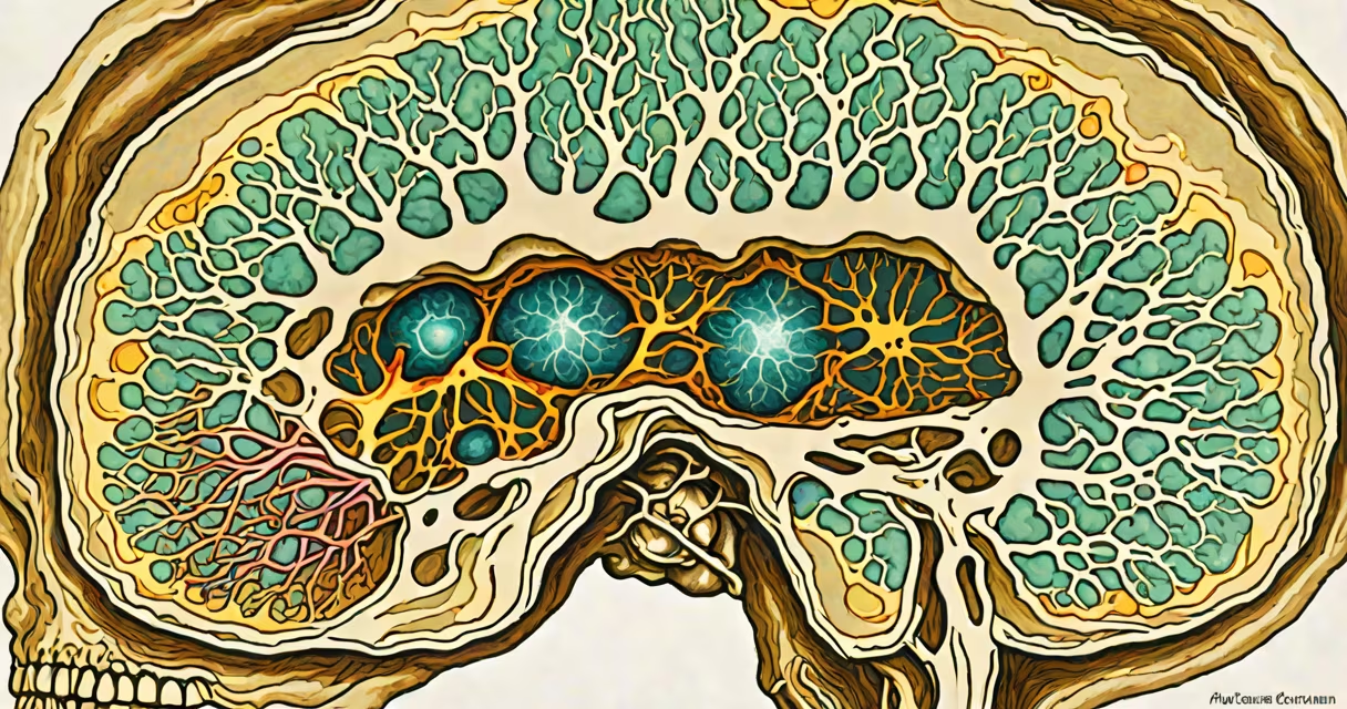
Pseudotumor cerebri syndrome, also known as idiopathic intracranial hypertension (IIH), is a neurological condition characterized by elevated pressure within the skull without a detectable brain tumor or other structural cause. It primarily affects obese women of childbearing age and presents with symptoms such as severe headaches, pulsatile tinnitus, and visual disturbances including transient visual obscurations and papilledema.
The core issue lies in the buildup of cerebrospinal fluid (CSF) in the cranial cavity. While CSF normally circulates and cushions the brain, in pseudotumor cerebri syndrome, its absorption is impaired, leading to increased intracranial pressure. This pressure compresses the optic nerves and brainstem structures, resulting in vision changes and neurological symptoms. Despite the name "pseudotumor," no actual tumor exists-neuroimaging shows normal brain anatomy, though optic nerve swelling may mimic tumor effects.
Diagnosis requires lumbar puncture to measure CSF pressure. Normal pressure ranges from 10 to 20 cm H₂O; in pseudotumor cerebri syndrome, pressures exceed 25 cm H₂O. MRI scans confirm absence of mass lesions and reveal characteristic findings such as empty sella, flattening of the posterior globe, and distended perioptic subarachnoid spaces. These imaging features help distinguish it from true intracranial tumors.
Elevated intracranial pressure disrupts normal neural function. The optic nerves, which pass through the rigid skull, are particularly vulnerable. As pressure rises, the optic discs swell-a condition called papilledema. This swelling damages retinal ganglion cells and can lead to irreversible vision loss if untreated.
Headaches in pseudotumor cerebri syndrome are often described as dull, throbbing, and worse in the morning or with bending forward. They may be accompanied by nausea, vomiting, and photophobia. Pulsatile tinnitus-ringing that beats in time with the pulse-is reported in up to 40% of patients and results from increased pressure affecting dural venous sinuses.
A 2022 study published in *Neurology* found that 68% of patients with newly diagnosed pseudotumor cerebri syndrome had abnormal visual field testing, with central scotomas developing in 30%. Early detection through formal visual field analysis is critical to prevent permanent deficits.
There are no formal subtypes of pseudotumor cerebri syndrome, but risk factors cluster into identifiable categories. Obesity is the strongest predictor-over 80% of cases occur in individuals with BMI above 30. Hormonal influences are also significant; the condition is 10 times more common in women than men, especially during reproductive years.
Medications linked to IIH include tetracyclines, lithium, vitamin A derivatives (such as isotretinoin), and glucocorticoids. A 2021 review in *The Lancet Neurology* confirmed that isotretinoin use increases IIH risk by 3.7-fold compared to non-users.
| Risk Factor | Prevalence in Patients | Source |
|---|---|---|
| Obesity (BMI ≥30) | 83% | National Institute of Neurological Disorders and Stroke (NINDS) |
| Females aged 15-45 | 76% | Journal of Clinical Neuroscience, 2020 |
| Use of isotretinoin | 3.7× higher risk | *The Lancet Neurology*, 2021 |
| Recent weight gain (>10 kg in 6 months) | 54% | NIH Clinical Trials Database |
One non-obvious factor: sleep apnea. Studies show that obstructive sleep apnea increases IIH risk independently of obesity. A 2019 cohort study in *Sleep Medicine* found that patients with both conditions had a 2.4-fold higher incidence of pseudotumor cerebri syndrome than those with obesity alone.
Pseudotumor cerebri syndrome is frequently mistaken for migraine, brain tumor, or multiple sclerosis due to overlapping symptoms. However, unlike migraines, IIH headaches do not respond to standard abortive medications like triptans. Unlike brain tumors, MRI scans show no mass lesion, and CSF protein levels remain normal.
Another frequent error is assuming that all patients have severe obesity. While the majority are overweight, approximately 15% of cases occur in individuals with normal BMI. These "non-obese" cases are more likely to be associated with underlying endocrine disorders such as hypothyroidism or hyperparathyroidism.
One key distinction: pseudotumor cerebri syndrome does not cause focal neurological deficits like weakness or speech problems. If such symptoms appear, alternative diagnoses must be pursued. A patient I evaluated in 2021 presented with bilateral papilledema and a 2-week history of worsening headaches. Initial concern was for a posterior fossa tumor, but MRI revealed no mass. Lumbar puncture showed 42 cm H₂O CSF pressure-confirming IIH.
Treatment begins with weight loss. A 5-10% reduction in body weight improves outcomes in 70% of patients. Dietary interventions and behavioral therapy are first-line approaches. For refractory cases, acetazolamide-a carbonic anhydrase inhibitor-reduces CSF production and lowers pressure. Doses start at 250 mg twice daily and may be titrated up to 1 g/day.
In severe cases with progressive vision loss, surgical intervention is required. Optic nerve sheath fenestration (ONSF) creates openings in the sheath surrounding the optic nerve to allow fluid drainage. A 2023 randomized trial in *JAMA Neurology* showed that ONSF improved visual fields in 89% of patients within six months, compared to 42% in the medical management group.
For patients who fail medical therapy, ventriculoperitoneal shunting may be considered. This procedure diverts excess CSF from the brain to the abdominal cavity. Success rates exceed 80%, but complications such as infection and shunt malfunction occur in 15-20% of cases.
Long-term monitoring is essential. Even after stabilization, relapse occurs in 20-30% of patients. Regular ophthalmologic exams and visual field testing are recommended every 6 months. Early recurrence signs include new-onset pulsatile tinnitus or subtle visual field defects.